Left/right asymmetry in vertebrates
The body plan of organisms undergoing embryogenesis occurs along three orthogonal axes; Anterior–posterior (A-P), dorsal–ventral (D-V) and left–right (L-R) axes. While the development of A-P and D-V axes has been well characterized, the molecular controls of the process in the L/R asymmetry is not yet fully understood.
On the surface, vertebrate organisms appear to be symmetrical (bilateral symmetry). However, the positioning of the internal organs are not symmetrical; the left and right halves of the body are not mirror images of one another. It can be seen in organs of the thoracic cavity such as the cardiovascular system, organs in the abdominal cavity (e.g gut, liver), as well as in the brain. This asymmetry is of great importance on morphological and/or functionality of various organs, thus maintaining a healthy development and everyday functioning of the organism. However, there are individuals that are developed to term with a partial or complete mirroring of the organs (situs inversus, as opposed to situs solitus which is the normal development).
The heart is the first organ to show visible asymmetry. During embryogenesis the heart starts out as a simple tube that eventually loops to the right. (It ultimately locates the heart in the center of the chest, with the muscle mass being greater on the left side). Meanwhile, the other organs move to their respective locations. The stomach, for instance, moves 90 degrees clockwise around a longitudinal axis. The dorsal wall grows faster than the ventral wall, causing the greater curvature to be located on the left side. Simultaneously liver growth is mainly situated to the right from the midline. Even symmetrically paired organs, such as the lungs, show asymmetry in their different number of lobes. An interesting scientific concept is how an otherwise bilaterally symmetric embryo can differentiate left from right, and organize it’s organs accordingly. The breakage of symmetry occurs prior to the visible morphological asymmetries of the developing organs, (Babu D, Roy S. 2013)
The establishment of L/R asymmetry
The mechanism of L/R asymmetry can be divided into 4 main steps
- Initial breaking of L/R symmetry in or near the node.
- Transfer of L/R signals from the node to the lateral plate mesoderm (LPM).
- L/R asymmetric expression of signaling molecules, such as transforming growth factor beta related molecules to e.g. Nodal, Lefty on the left side.
- L/R asymmetry morphogenesis of the visceral organs that are induced by these signals.
Nodal Signaling
Nodal signaling plays an essential role in early embryogenic cell differentiation, formation of mesoderm and ectoderm, and development left-right axial structures. Evidence for the latter derive from genetic- and surgical manipulation of fish-, amphibia-, chick-, and mammalian embryos. Nodal signaling is regulated by several mechanisms; ligand processing, co-factors, antagonists and positive- and negative feedback loops.
Node
The notochord, formed during gastrulation, is an embryonic midline structure facilitating several important mechanisms during embryogenesis. It secretes signaling factors to all surrounding tissues, giving information on positioning and fate of various organs. This makes it an important contributor to formation of L/R-asymmetry. Located at the end of the notochord is an embryonic cavity, the node. The node secretes many important proteins such as Nodal, Lefty2 and Pitx2.
Nodal, Lefty & Pitx2
Nodal is a protein ligand of the Transforming Growth Factor (TGF-beta) superfamily. On the surface of cells, Nodal can bind to heterodimer receptors, consequently activating the cells. The activation causes the phosphorylation of Smad2 or Smad3 proteins. Smad2/3 then binds with Smad4 creating a smad complex. This complex can migrate to the nucleus and act as an transcription factor, promoting expression of target genes (including its own) involved in cell differentiation and proliferation.
Cells that receive nodal signaling will adopt left sided morphology, the ones that does not will adopt right sided morphology. Lefty is also a protein of the TGFB superfamily. They antagonise nodal signaling by blocking formation of receptor complexes. Lefty is of great importance in L/R specification because it repeatedly act as a downstream target of Nodal, causing a negative-feedback mechanism for nodal signaling. Pitx2 can be activated by Nodal, causing left-sided morphogenesis in the lateral plate mesoderm (LPM). Firstly Nodal is initiated by an unknown mechanism (see later), generating an expression of the protein exclusively on the left side of the LPM. Here Nodal activates Lefty, where they both have asymmetric enhancers (ASE) which is crucial to the left-sided morphogenesis. (Hiroshi Hamada, Chikara Meno et al. February 2002)
Nodal autoregulates itself in a positive feedback loop, while Lefty is an antagonist of Nodal, restricting the range and duration of the signaling pathway. There is a balance of activation and inhibition, which is fine tuned to accomplish precise expression of the various genes important in early embryogenesis. (Hiroshi Hamada, Chikara Meno et al. February 2002) Because Lefty2 is an monomer, and Nodal is an dimer, it can diffuse in a longer range. In this way Lefty can inhibit Nodal on the right side of the LPM as well. Only cells in close proximity to nodal is activated. (Babu D, Roy S. 2013)
Initial left-right symmetry breakage
The works of Nodal, Lefty2 and several other factors contributing L/R-asymmetry has been well researched in several different species, however knowledge of what causes the initial breakage of symmetry still an on-going discussion today.
Theories
Two models are currently thought to be mutually plausible. Leftward flow of extracellular fluid caused by Cilia, and asymmetric signals generated by an ion flux in the zygote during the first cleavage divisions. The latter theory also states that cilia-driven flow of EC fluid merely has a later-stage supporting effect.
Leftward flow caused by Cilia
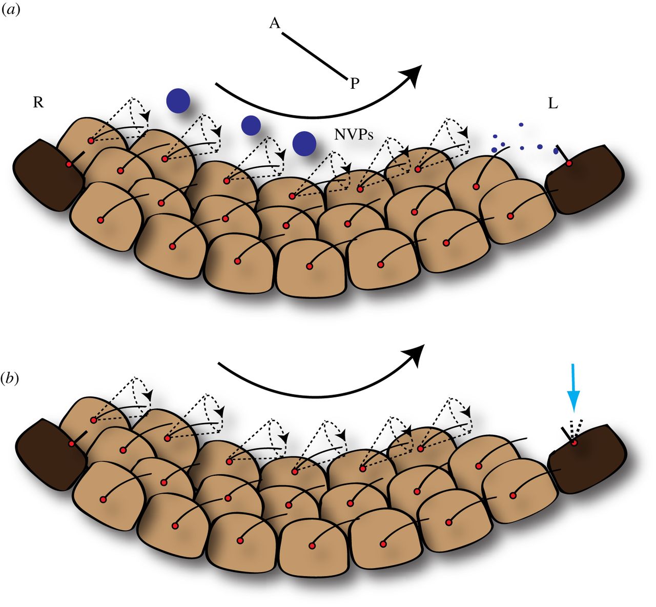
A heavily supported theory is the leftward flow of extracellular fluid caused by Cilia. The fluid flow cause a different degree of signaling molecules on the left vs the right.
Cilia are hair-like projections found on the surface of cells. There are two types of cilia: motile cilia, longer and has the ability to beat, generating a fluid-flow non-motile cilia, transduct sensory signals
The influence cilia has on L/R-asymmetry was discovered because of a rare human condition; Kartagener syndrome. This syndrome causes loss of motility in respiratory cilia and sperm flagellum, and situs inversus (Inversion of the organs). (Hiroshi Hamada, Chikara Meno et al. February 2002).
The interior of a tiny node cavity on the embryo’s midline is lined with 200-300 cilia which rotate at a rate of 10 times a second. Quantitative analysis show that L/R - asymmetry is determined already within a few hours after the beginning of fluid flow. Only a weak flow will start an embryo on its proper L/R maturation, and only two cilia is necessary to generate this flow. (K. Shinohara, A. Kawasumi et al. 2012) The cilia are tilted, therefore creating a flow in one specific direction, from right to left. The tilt is crucial for their ability to organize the early developing embryo into left and right. Interestingly, scientists have disabled the genes necessary for the cilia to tilt, creating a mutation causing a mouse embryo to develop situs inversus. (Babu D, Roy S. 2013) The clockwise rotational movement cause the fluid flow to reach the left side of the rim of the node. Although there are some uncertainties concerning the exact mechanics, the non spinning cilia at the rim are triggered by the initial flow - either by a physical bend or possibly by released calcium ions that spread to surrounding cells . The cells secrete Nodal, creating a feedback loop causing other cells to do the same. (Babu D, Roy S. 2013)
|
Ion flux theory
The ion flux theory suggest that the initial breaking of symmetry happens already in the first cell division of the zygote. It’s proposed that a maternal ion pump (ATP4) is deposited by motor proteins. The ion pump will generate an intracellular pH and voltage gradient, fueling the transfer of serotonin via gap junctions between the cells. The serotonin will be unequally distributed; inhibiting nodal activity on the right side, thus causing asymmetric activation of the nodal signaling pathway. Furthermore this theory state that nodal flow caused by cilia is only maintains L/R - asymmetric development at a later stage in embryogenesis.

 |
|