The Thyroid: Congenital hypothyroidism in Foals
By Andrew Cachia, Andrea Grima & Aoife Kingston
Contents
1.0 What is congenital hypothyroidism and what are its symptoms?
Congenital hypothyroidism is a disease which occurs naturally, and is caused by the hyperplasia1 of the thyroid gland. This alters the ability of the thyroid gland to uptake iodine either causing too much iodine to be present, or too little. This is regulated by the thyroid stimulating hormone, which is controlled through negative feedback by the two endocrine hormones; triiodothyronine-T3 and tetraiodothyronine also known as thyroxine - T4. Congenital hypothyroidism leads to musculoskeletal deformities in newborn foals, and was one of the main causes of foal mortality during the 1990’s (Washington state college, (2006) ) which mainly affecting the northern parts of North America.
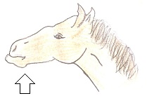
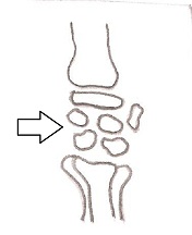
Many experiments and analysis were made, in order to understand what the naturally occurring disease is, and to expose its pathogenic background. It was observed that affected foals were typically born between 340 and 400 days after conception, making the gestation period more than a month longer than that of a healthy foal. These foals were diagnosed as suffering from hypothyroidism, and were born with various musculoskeletal deformities. Very often these deformities consisted of the ruptured tendons, limbs and mandibular progmathism2, with the most common deformity being the incomplete ossification of the carpal and tarsal bones. Figure 1 displays mandibular prognathism, whilst figure 2 highlights the incomplete ossification in the carpus of a hypothyroid foal.
 Figure 1
  Figure 2
During the course of the investigations, some healthy foals had their thyroids removed 210 days into the gestation whilst still in the womb. These foals also showed the same lesions and deformities as would be found in naturally occurring hypothyroid foals. These findings confirmed that the foals which naturally contracted the disease were hypothyroid whilst in the uterus, therefore, the lesions and deformities present were due to the disease at hand, and not caused by some other underlying condition. There are two main types of congenital hypothyroidism which are caused by two different phenomena; the first type is the congenital hypothyroidism due to the insufficient iodine presence during the gestation period. The second kind of congenital hypothyroidism is caused by excess iodine presence during the gestation period (Allen, 1995) (Washington state college, 2006)
1.1 The importance of T3 and T4
Through the action of TSH3 which is produced in the pituitary gland. The follicular cells present in the thyroid gland absorb iodine at rates which depend on the secretion levels of the TSH. After absorbing the iodine,the follicular cells convert it into tyrosine molecules to produce the two hormones; triiodothyronine (T3) and tetraiodotyronine, also known as thyroxine (T4). The numbers following the T, indicate the number of iodine particles the molecule can carry. T3 has the ability to carry three Iodine particles, whilst T4 has the ability to carry four Iodine particles.
After secretion, the T3 and T4 molecules are released into the blood, and follow in the blood circulation reaching cells throughout the body, and being absorbed as they move along. On absorption into a cell, T4 is converted into T3 by the removal of one of the Iodine particles. T3 is the active form of the two hormones, which is the active ingredient that brings about the changes which can be observed in the body as a result of the thyroid hormones. Even though T4 is simply converted into T3, it may seem as if it has a less significant role in the body. However, it has an important function which provides negative feedback to the pituitary and hypothalamus glands, to regulate the secretion of the thyroid stimulating hormone.
Due to the two hormones being the main regulators of the body’s metabolism and energy synthesis, the absence, of reduced concentration of the two would lead to a low basal metabolic rate, which leads to hypothyroidism. Signs of hypothyroidism in adult horses include hair loss, lethargy and weight gain even though a normal level of food consumption is observed. If a foal is born to a mare suffering from this condition, then it would be born with deformities which correspond with an animal suffering from hypothyroidism. (Washington state college, 2006) (B.C McLaughlin, C.E Doige and P.S McLaughlin, 1986) (Allen, 1995)
1.2 Factors affecting thyroid hormones
Age: There is a known phenomenon that when it comes to neonatal foals in the mare’s womb, their thyroid hormone levels are very high when compared to that of adult horses. Within the first 48 hours after birth, T3 levels are typically between 6-15mmol/L whilst T4 levels are usually between 200 and 600 mmol/l. After the first week, the levels of both hormones begin to decline considerably, but remain remarkably high in comparison to those of adults till about one year of age. Normal level of T4 are indicated as being between 279-464 mmol/l, while the figure for T3 is When horses exceed the one year benchmark, there is no real difference between the T3 and T4 levels of a one year old horse and a ten year old horse. (Nat T.M., Thomas R., Josie L.T., David A.K.J., Donald L.T, 1998)
Breed: Very little studies have been carried out to investigate the correlation between the breed of the foal, and its relation to foals suffering from hypothyroidism. However, the little work done has established that there is no correlation between the breed of the animal and hypothyroidism.
Sex: in the case of the gender of the animal, it was previously thought that no real relationship was present between the gender and the thyroid hormone secretions. However, after preliminary studies it was found that stallions tend to have higher concentrations of T4 than those found in mares and foals.
Reproductive status: it is known that the concentrations of the T3 and T4 hormones in mares alter with very minimal altercations. Through various studies it has been found that, the T4 concentrations in mares tend to decrease slightly once ovulation has occurs. It was also found that when a mare becomes pregnant, the T4 levels are the same as those found in non pregnant females. However, in the case of the T3 hormone, the levels decrease steadily throughout the pregnancy. The final relationship found between the two is that in lactating mares, the T4 concentrations were much lower when compared to mares which weren’t lactating. (Allen, 1995)
2.0 Iodine
75% of Iodine in the body is located in the thyroid gland. It is an essential nutrient for reproduction and normal physiological function in the horse. Insufficient or excess iodine may lead to congenital hypothyroidism in the foal, as it affects the levels of T3 and T4 circulating in the blood.
2.1 Excess iodine in diet of mare during gestation
Excess iodine in the diet of the Mare may lead to Hypothyroidism in the foal. Mares supplemented with more than 35mg/day of iodine may produce affected foals. Iodine is highly concentrated across the placenta and in the milk, therefore the fetus and new born foal receive much higher concentrations of iodine than that present in the mare’s feed.
A study found that the occurrence of goiter was proportional to the level of iodine being fed to the mares. 3% of foals on one farm feeding 48-55 mg I/day, 10% on a farm feeding 36-69 mg I/day and 50% on another farm feeding 288-432 mg I/day. (Frederick H., Warren G, 2006)
2.2 Dietary Iodine
There are certain food groups that are much higher in iodine than others. One example is Kelp, a specific family of seaweeds. It can have as much as 1850 ppm of iodine, while the required iodine intake is estimated to be 0.1 ppm4 or 1-2 mg per day. (Frederick H., Warren G, 2006) Certain plants, when ingested in sufficient amount, may cause goiter in the mare, as they contain goitrogenic substances5 These materials can be removed through cooking or heating. The Soybean is the most notable, but other examples which are less potent are Cabbage, Rape and Kale. These goitrogenic substances are not usually significant in the mare. However, hypothyroidism may develop in the foal as a result of the ingestion of these materials.
2.3 Iodine deficiency in mares diet during gestation
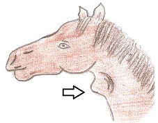
Iodine deficiency reduces the ability of the thyroid gland to produce thyroid hormone. Excessive plasma iodine levels inhibits iodine uptake by the thyroid gland, thyroidal peroxidase activity and thyroxin synthesis. This leads to reduced levels of thyroid hormones being circulated in the body. Any disruption to the hypothalamic-Pituitary-Thyroid axis results in such reduced levels. This causes the pituitary to secrete more TSH which leads to hyperplasia of the thyroid gland and results in goiter. The Hyperplastic Gland may compensate, and often does, for the reduced iodine level therefore Hypothyroidism is in no way the same thing as goiter. However, foals born to mares that are insufficient in iodine are likely to have thyroid enlargement and clinical signs of hypothyroidism, as can be seen in figure 3 which shows an enlarged thyroid in a foal suffering form goiter.
 Figure 3
2.4 Nitrate Levels of Forage
High levels of nitrates have also been found to impair the function of the thyroid gland. A Study carried out it Canada illustrated that congenital hypothyroidism and dysmaturity syndrome<<footnote(Foals are born after their expected due date, however are small and have the characteristics of being premature)>> in foals may be caused by high nitrate levels in the diet of the mare (Allen, 1996). A case control study of the congenital hypothyroidism and dysmaturity syndrome in foals, found that the chance of a foal being born with the disease on a farm that fed forage with at least a trace of nitrate, was 8 times higher than the chance that the disease would occur on a farm that fed forage free of nitrate. Furthermore, the chance of a mare producing a foal with Hypothyroidism is 5.9 times greater than that of a mare being fed nitrate free forage. It also showed that Mares being fed green feed without any supplements had a higher risk of producing a foal with the disease. (Allen, 1996)
3.0 Musculoskeletal Deformities
Thyroid hormones play a vital role in the early growth and development stages of the fetus during pregnancy. Musculoskeletal deformity6 is a syndrome characterised by the decreased function of the thyroid gland in the developing fetus. This disorder is undetected during the gestation period, however, once the foal is born the full extent of this iodine deficiency, or overdose is apparent. This syndrome is well recognised in western Canada, where low levels of iodine in green feed continues to be an important cause of reproductive loss and foal mortality. (Allen, 1995)
Although the mare may have given birth on schedule, or up to a month late, the foal would have a premature appearance. The foal would have a soft silky coat, droopy ears, unusually pliable hooves, poor muscle development, poor suckling reflexes and lax tendons and joints. These abnormalities hinder the foal, in that it would have difficulty standing and nursing on its own. As a result, the foal’s prognosis would be very poor with the majority being euthanized.
Along with the latter greatly impacting the foal’s survival, the foal could also be born with multiple deformities which would also greatly decrease its chances of survival. Such deformities include mandibular prognathism , poorly or incompletely ossified carpal and tarsal bones, ruptured tendons of the common digital extensor muscle and doming of the head. In figure 4 the degree of hyperplasia of the cuboidal bones of the carpus is visible, featuring the comparison between a severely deformed to a normal carpus. The carpus is affected most frequently, but the tarsus and fetlocks are occasionally involved. The deviation is obvious but varies in severity. A valgus7 of up to 6° of the distal portion of a limb may be regarded as normal. Outward rotation of the fetlocks invariably accompanies carpal valgus. Foals with defective ossification of the carpal cuboidal bones or excessive joint laxity are frequently lame as the legs become progressively deviated. Affected limbs of the foal must be palpated carefully to detect ligament laxity and specific areas that may be painful. (Briggs K. 2011)
Figure 4 
3.1 Study on T3 and T4 serum levels of foals suffering from musculoskeletal deformities
As mentioned earlier T3 and T4 play an important role in the muscular and skeletal growth and development of the foal during pregnancy. Therefore, a deficiency in these thyroid hormones may impact the foal’s development. A study carried out by B.G McLaulin, C.E Doige and P.S McLaulin on fourteen foals with congenital musculoskeletal abnormalities, found a correlation between low serum levels of T3 and T4 and hypothyroidism. Serum samples were assayed for total serum T3 and total serum T4 by radioimmunoassay(RIA8). In selected cases affected limbs were radiographed. Seven Severely affected foals were euthanized and complete necropsy preformed. Selected tissues were collected from these subjects for histological analysis.
Historical Date Lesions and Thyroid Hormone Levels Fourteen Foals
Foal |
Breed |
Sex |
Age |
Thyroid |
Musculoskeletal deformities |
T3(nmol/L) |
T4(nmol/L) |
1 |
Appaloosa |
Male |
1 day |
Goiter |
F.R.M |
2.2(Low) |
136.4(Low |
2 |
Quarter horse |
Female |
1 day |
Not examined |
A.F.R.M.S |
2.5(Low) |
95.2(Low) |
3 |
Arab |
Male |
3 days |
Not examined |
M |
9.0(Low) |
101.7(Low) |
4 |
Quarter horse |
Male |
4 days |
Not examined |
A.F.S |
1.6(Low) |
25.7(Low) |
5 |
Quarter horse |
Male |
7 days |
Goiter |
F.R.M |
3.3(Low) |
117.0(normal |
6 |
Quarter horse |
Unknown |
7 days |
Not examined |
F.M |
7.1(Low) |
120.0(normal) |
7 |
Quarter horse |
Male |
7 days |
Not examined |
A |
7.6(Low) |
294.0(normal) |
8 |
Quarter horse |
Male |
10 days |
Not examined |
R |
8.2(Low) |
140.0(normal) |
9 |
Appaloosa |
Female |
10 days |
Goiter |
A |
1.8(Low) |
217.5(normal) |
10 |
Thoroughbred |
Female |
21 days |
Not examined |
R |
1.4(Low) |
18.0(normal) |
11 |
Standard bred |
Female |
1 month |
Goiter |
A.F.R.S |
1.1(Low) |
3.9(Low) |
12 |
Quarter horse |
Female |
35 days |
Goiter |
A.S |
4.1(Low) |
52.8(normal) |
13 |
Thoroughbred |
Male |
3 months |
Not examined |
S |
0.7(Low) |
12.9(Low) |
14 |
Quarter horse cross |
Male |
5 months |
Not examined |
A.S |
5.1(Low) |
0.9-1.5(Low) |
A |
Angular limb deformity |
F |
Forelimb contraction |
R |
Ruptured common digital extensor tendon |
S |
Skeletal hypoplasia |
M |
Mandibular prognathism |
From the table it can be understood that more foals had lower serum levels of T3 and T4 in their blood, indicating that they were suffering from hypothyroidism. 85% of the examined euthanized foals were documented as having goiter, which further strongly suggests that there is a direct link to musculoskeletal deformities and hypothyroidism.
This study described the foals as being weak at birth, and requiring assistance to suckle. The foals with forelimb contortions were unable to stand without assistance. In all cases musculoskeletal lesions were detected shortly after birth. Angular deformities were present as lateral or medial deviations originating at the carpus or tarsus, which were incompletely ossified and showed a thickening layer of articular cartilage. The affected carpal bones showed deformities, and the third tarsal bones were collapsed. The forelimbs were in fixed flexion, and the fetlocks could not be straightened. Rapture of the common digital extensor tendon was often present, also the foals were observed to have mandibular prognathism. Examination of the thyroid gland of euthanized foals showed normal size and shape, however, the thyroid appeared paler and firmer than normal, which further indicates that the foals were suffering from goiter.(B.C McLaughlin, C.E Doige and P.S McLaughlin, 1986) */
4.0 Prognosis and Treatment
Many foals with congenital hypothyroidism go through prolonged gestation periods, yet are poorly developed and immature. The prognosis of the foal mainly depends on their level of immaturity. However, it is generally very poor, as many would either be still born, aborted or may even need to be euthanized shortly after birth.
The Mandibular prognathism usually corrects itself over time. Splints or tube casts are often used for underdeveloped bones, as a means of support while standing. They also help the bones grow correctly, without damaging the cartilage and soft bones. Exercise should also be restricted until the bones develop further and look stronger on the x-ray (Briggs K.(2011)). Many affected foals have respiratory problems, due to their poorly compliant lungs and greatly compliant chest (Beers M. H. (2011)). These foals would need to be given oxygen through a nasal tube until the reparatory organs have matured.
Hypothyroidism is commonly treated with thyroid drugs as hormone supplements. Synthetic levothyroxine (T4) and synthetic liothyronine (T3) are commonly used. Synthetic T4 is the most popular drug used, as its greatest advantage is that it allows each tissue to individually convert T4 to T3 to meet its metabolic need. It is also able to trigger the normal negative feedback mechanism, so that it closely imitates the normal regulatory mechanism for hormone production. The disadvantage to using only T3 supplementation is that it supplies the same level of hormone to each of the tissues, as it bypasses the local tissue regulation of thyroid hormone conversion. This explains why T4 supplementation is more widely used.
Thyroid supplementation is generally given once daily, and T4 is administrated according to blood T4 concentrations. Thyroid extracts are no longer commonly used, however, they are still available for use by some Veterinary Surgeons. They are made from bovine or porcine thyroid glands which are acquired from slaughter houses. The effectiveness of these extract is based on the level of iodine they contain rather than the level of T3 or T4. (Clinical Pharmacology and therapeutics for the veterinary technician)
5.0 References I
Allen A.L., Townsend H.G., Doige C.E., Fretz P.B. (1996): A case-control study of the congenital hypothyroidism and dysmaturity syndrome of foals. The Canadian Veterinary Journal, 37: (6) 349-358, http://www.ncbi.nlm.nih.gov/pmc/articles/PMC1576403/
Allen A.L. (1995): Hyperplasia of the thyroid gland and musculoskeletal deformities in two equine abortuses. The Canadian veterinary Journal, 36: (4) 234–236, http://www.ncbi.nlm.nih.gov/pmc/articles/PMC1686925/?page=1
Frank N., Sojka J.E., Latour M.A.(2004): Effect of hypothyroidism on the blood lipid response to higher dietary fat intake in mares. Animal Science Journal, 82: (9) 2640-2646, http://www.journalofanimalscience.org/content/82/9/2640.full.pdf+html?sid=7466fdd7-527c-4e81-ad0d-d4052afb794a
Frederick H., Warren G.(2006):Minerals for Horses, Part II Trace Minerals. Horse Express Journal, 25: (1) http://www.amavitahorse.com/Frederick_Harper_article-express.pdf
Joe. D.P.(2011): Micromineral requirements in horses. Kentucky Equine Research Journal, 317-328, http://www.ker.com/library/advances/239.pdf
Nat T.M., Thomas R., Josie L.T., David A.K.J., Donald L.T.(1998): Thyroid Hormone Levels in Thoroughbred Mares and Their Foals at Parturition. Medicine II Journal, 44: 248-251, http://www.ivis.org/proceedings/aaep/1998/messer.pdf
B. G. McLaughlin, C. E. Doige, P. S. McLaughlin (1986), Thyroid Hormone Levels in Foals with congenital musculoskeletal lesions, Canadian Veteriinary Journal, 27: (7), 246-267, http://www.ncbi.nlm.nih.gov/pmc/articles/PMC1680276/?page=1
Kreplin C., Allen A. (1992), Congenital hypothyroidism in foals in alberta, Canadian Vet Journal, Vol 32, 751, http://pubmedcentralcanada.ca/picrender.cgi?accid=PMC1481118&blobtype=pdf
References II
Briggs K.(2011): Congenital Hypothyroidism in foals, 61-64, http://ridexc.files.wordpress.com/2011/11/premature-foal.pdf
Beers M. H.(2011) : Prematurity, Dysmaturity, and Postmaturity. The Merck Veterinary Manual, http://www.merckvetmanual.com/mvm/index.jsp?cfile=htm/bc/160820.htm
Kevin D.D.(1998): THYROID HORMONES AND THEIR EFFECT ON REPRODUCTIVE FUNCTION, 1-2, http://www.equinereproduction.com/articles/pdf/THYROID%20HORMONES%20AND%20THEIR%20EFFECT%20OF%20REPRODUCTIVE%20FUNCTION%20_Original%20Print-1998_.pdf
Munroe G.(2012): Endocrine hypothyroidism. Vetstream, 2-8, http://www.vetstream.com/equis/Content/Disease/dis00312
Palmer J.E.: Prematurity, Dysmaturity, Postmaturity. Neonatology Journal. http://nicuvet.com/nicuvet/Equine-Perinatoloy/NICU%20Lectures/Prematurity.pdf
Washington state college(2006): Veterinary Hypthyroidism in foals. Equine news Journal, Vol 3, No. 2 http://www.vetmed.wsu.edu/depts-vth/EquineNews/archive/112881EquineNewsSp06color.pdf
IODINE IN THE HORSE TOO MUCH OR TOO LITTLE, 2002 http://www.4source.com/technical/iodine1.shtml
TSH. Lab Tests Online, 2012 http://labtestsonline.org/understanding/analytes/tsh/tab/test
Clinical Pharmacology and therapeutics for the veterinary technician Journal,In Drugs affecting the endocrine system, (7) http://www.vet.purdue.edu/vtdl/bms_235/PDFbookchaps/Chapter%207%20-%203rd%20ed%20-%20endocrine.pdf
Beers M. H.: Non-neoplastic enlargement of the thyroid gland, The Merke Veterinary Manual http://www.merckvetmanual.com/mvm/index.jsp?cfile=htm/bc/40603.htm
Investigation into congenital hypothyroidism of foals, A Thesis http://library.usask.ca/theses/available/etd-10212004-000356/unrestricted/nq24002.pdf
Beers M. H.: Hypothyroidism, The Merke Veterinary Manual http://www.merckvetmanual.com/mvm/index.jsp?cfile=htm/bc/40602.htm
References III
http://www.vetnext.com/search.php?s=aandoening&id=73059460610%20431
http://onlinelibrary.wiley.com/doi/10.1111/j.2042-3306.1984.tb01932.x/abstract
Figures:
Figure 1 Dr Siddra Hines, Emmy Widman,(2010) Facts and Myths about equine hypothyroidism, : http://www.google.com/imgres?um=1&hl=en&sa=N&tbo=d&biw=2051&bih=965&tbm=isch&tbnid=IMH6bEOaTjjWDM:&imgrefurl=http://www.equinechronicle.com/health/facts-and-myths-about-equine-hypothyroidism.html&docid=67VNbaCeCcUOgM&imgurl=http://www.equinechronicle.com/wp-content/plugins/fresh-page/thirdparty/phpthumb/phpThumb.php%253Fw%253D419%2526src%253D/home/equinech/public_html/wp-content/files_flutter/1272575606hypothyroid.jpg&w=418&h=314&ei=w6ijUKruCInMsgaVwYCADw&zoom=1&iact=hc&vpx=5&vpy=152&dur=2154&hovh=195&hovw=258&tx=103&ty=113&sig=106250435140341025680&page=1&tbnh=138&tbnw=178&start=0&ndsp=65&ved=1t:429,r:0,s:0,i:74
Figure 2; shows the incomplete ossification of a carpus in a hypothyroid foal redrawn based on the figure of: http://www.google.com/imgres?um=1&hl=en&sa=N&tbo=d&biw=2051&bih=965&tbm=isch&tbnid=qu9M_UJKMDiVYM:&imgrefurl=http://www.thehorse.com/images/content/congenital_hypothyroidism/congenital_hypothyroidism.html&docid=nP9d8XFf_QtMWM&imgurl=http://www.thehorse.com/images/content/congenital_hypothyroidism/abnormal_carpus.jpg&w=260&h=387&ei=9q2jUJ7xDNHMtAbKiYCYBA&zoom=1&iact=hc&vpx=182&vpy=119&dur=114&hovh=273&hovw=184&tx=85&ty=155&sig=106250435140341025680&page=1&tbnh=151&tbnw=96&start=0&ndsp=67&ved=1t:429,r:1,s:0,i:77
Figure 3 Enlarged thyroid, Foal redrawn based on the figure of: http://www.merckvetmanual.com/mvm/htm/bc/endth01.htm
Figure 4 Severe and Mild case of hyperplasie of carpus and normal cuboidal bones of the carpus of a foal redrawn based on the figure of: http://ridexc.files.wordpress.com/2011/11/premature-foal.pdf
Table 1 B. G. McLaughlin, C. E. Doige, P. S. McLaughlin(1986), Thyroid hormone levels in foals with Congenital musculoskeletal lesions, Canadian Veterinary Journal, 27(7) 264-266,267,: http://www.ncbi.nlm.nih.gov/pmc/articles/PMC1680276/?page=2
The increase in the number of cells of a particular tissue which can lead to the enlargement of an organ (1)
The mandible is longer than the maxilla (2)
Thyroid stimulating hormone (3)
Parts per million, which is a measurement of dilute concentrations. (4)
Substances that interfere with thyroid function, causing goiter. (5)
Foals that are born after their expected due date, however are small and have the characteristics of being premature (6)
The lateral deviation of a bone or joint. (7)
Technique used to measure concentration of antigens (8)
