|
Size: 22053
Comment:
|
← Revision 81 as of 2017-05-03 16:54:00 ⇥
Size: 22027
Comment:
|
| Deletions are marked like this. | Additions are marked like this. |
| Line 101: | Line 101: |
| * Berken, Jonathan A., Vincent L. Gracco, and Denise Klein. "Early Bilingualism, Language Attainment, And Brain Development". Neuropsychologia 98 (2017): 220-227. Web. * Jens Brauer, Alfred Anwander, Daniela Perani, Angela D. Friederici. "Dorsal and Ventral Pathways in Language Development". Brain and Language 127.2 (2013): 289-295. Web. * Adam Felton, David Vazquez, Aurora I. Ramos-Numez, Maya R. Greene,Aleesandra Macbeth, Arturo E. Heranandez, Christine Chiarello. "Bilingualism Influences Structural Indices of Interhemispheric Organization". Journal of Neurolinguistics 42 (2017): 1-11. Web. * Friederici, Angela D. "The Cortical Language Circuit: From Auditory Perception to Sentence Comprehension". Trends in Cognitive Sciences 16.5 (2012): 262-268. Web. * Sini Hämäläinen, Viljami Sairanen, Alina Leminen, Minna Lehtonen. "Bilingualism Modulates the White Matter Structure of Language-Related Pathways". NeuroImage''' '''152 (2017): 249-257. Web. * Sawyer, Jeremy. "In What Language Do You Speak to Yourself? A Review of Private Speech and Bilingualism". Early Childhood Research Quarterly 36 (2016): 489-505. Web. |
* Berken, Jonathan A., Vincent L. Gracco, and Denise Klein. "Early Bilingualism, Language Attainment, And Brain Development". Neuropsychologia 98 (2017): 220-227. * Jens Brauer, Alfred Anwander, Daniela Perani, Angela D. Friederici. "Dorsal and Ventral Pathways in Language Development". Brain and Language 127.2 (2013): 289-295. * Adam Felton, David Vazquez, Aurora I. Ramos-Numez, Maya R. Greene,Aleesandra Macbeth, Arturo E. Heranandez, Christine Chiarello. "Bilingualism Influences Structural Indices of Interhemispheric Organization". Journal of Neurolinguistics 42 (2017): 1-11. * Friederici, Angela D. "The Cortical Language Circuit: From Auditory Perception to Sentence Comprehension". Trends in Cognitive Sciences 16.5 (2012): 262-268. * Sini Hämäläinen, Viljami Sairanen, Alina Leminen, Minna Lehtonen. "Bilingualism Modulates the White Matter Structure of Language-Related Pathways". NeuroImage''' '''152 (2017): 249-257. * Sawyer, Jeremy. "In What Language Do You Speak to Yourself? A Review of Private Speech and Bilingualism". Early Childhood Research Quarterly 36 (2016): 489-505. |
The Neural Background of Language Learning (Mother Tongue and Foreign languages)
Introduction
The neural background of language learning is still quite uncertain. There is no specific definition for language learning as it concerns too many areas. In simple terms, it can be defined as acquiring the ability to communicate in your mother tongue or a second/foreign language.
Recently focus has been put on bilingualism as more and more people are exposed to multiple languages. This is due to a greater exposure with technology, immigration and ethnic diversity.
Neuroplasticity is the reconfiguration of the brain due to environmental needs for “specific motor behaviour or cognitive skills”. These changes later have an influence on developing further competences but it seems that there is an optimal age to obtain skills even though neuroplasticity occurs at all ages (even at senescence).
The Method of language learning
Components involved in language learning
The brain regions that are used for language development can be found in the inferior frontal and temporal cortices, within these location dominance is seen in the left hemisphere.
It is here in these areas that we can find the main components of language learning and development, the ventral and dorsal pathways. The inferior frontal and temporal cortices are connected via these two pathways. The ventral pathways main purpose is for auditory-to-meaning mapping. This is the process in which the brain goes through in connecting the auditory sound of a word to its meaning. The dorsal pathway is needed for supporting auditory-to motor mapping. This is the association between of the sound of the word and the brain's skills used for articulatory purposes. Recently studies have shown that syntactic processing may also heavily rely on the dorsal pathway, in particular when sentences are complex (Friederici, Angela D. et al,2012).
The temporal cortex is connected to the premotor cortex by the dorsal pathway, which tracks through the inferior parietal cortex (IFC) and parts of the superior longitudinal fasciculus (SLF). The ventral pathway connects the temporal cortex to Brodmann Area (BA) 44, which is located in the frontal cortex as it is specific to humans and other primates, as part of Broca’s area via the arcuate fasciculus (AF). The ventral pathway also seems to be responsible for more than one function: it is assumed to support sound-to-meaning mapping, as well as local syntactic structure building or syntactic processes in general (Friederici, Angela D. et al,2012).
Currently, there are thought to be two functionally and structurally different dorsal pathways. There is also considered to be two ventral pathways in their possible relevance for semantic and syntactic processing during language comprehension (Friederici, Angela D. et al,2012).
Auditory perception is the initial stage in the auditory language comprehension process with this taking place in the temporal cortex. It is in the anterior region of Heschl's gurus that auditory processing takes place, this is in the left superior temporal gyrus. Here, we can see where a words phonological form or processing of the auditory word takes place.
The Dorsal Pathway
As mentioned above, the Dorsal pathway mainly involves the premotor cortex and the temporal cortex and with the inferior parietal cortex and the superior longitudinal fasciculus being passed through in the process.
With early age development data from diffusion weighted MRI, it is suggested that new-borns do not have a fully developed dorsal connection between the superior temporal gyrus to the inferior frontal gyrus. Researchers believe that the dorsal connection is terminated in the premotor cortex. With one connection terminating in the premotor cortex and the other in Brodmann area 44 of Broca’s Area, we can presume that this is the formation of two distinct dorsal pathways. The first dorsal connection to the Premotor cortex, D1, can be seen from birth. It also may be responsible for sensory-to-motor mapping which is necessary for early on language learning. The second dorsal pathway, D2, in the inferior frontal gyrus is not present at birth. Researchers are still uncertain as to how the two different pathways develop (Brauer, Jens et al. 2013).
Recent findings from developmental linguistics has shown that at the age of 7, the processing of complex sentences begins. In an experiment carried out by Perani et al (2011), it was proved that the D2 is present in children at the age of 7. However, we must recognise that even though the D2 pathway is complete in children, it is not fully matured. At this age, children are seen to successfully process passive sentence structures or object-first constructions. This may indicate that this dorsal pathway may be supported by Brodmann’s Area 44 (Brauer, Jens et al. 2013).
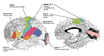
Figure 1. Brodmann's Area
The Ventral Pathway
The ventral pathway consists of two fibre tracts that run closely together, the uncinate fascicles and the extreme capsule fibre system.
The uncinate fasciculus, which connects the anterior ventral inferior frontal cortex to the temporal pole, and the extreme capsule fibre system, which mediates the inferior fronto-occipital fasciculus (IFOF), which connects the inferior frontal cortex along the temporal cortex to the occipital cortex.
The ventral pathway is split into two pathways, similar to the dorsal pathways, the superficial tract (V1) and the deep tract (V2). Scientists have suggested that the superficial tract is involved in the language network terminating in the pars triangularis and pars orbitalis of the inferior frontal gyrus. While the deep tract terminates in 3 frontal regions: An anterior component in the frontal pole and orbitofrontal cortex, a middle component in the middle frontal gyrus and a posterior component in the middle frontal gyrus and the dorsolateral prefrontal cortex (Brauer, Jens et al. 2013).
The deep tract of the inferior fronto-occipital fasciculus passes through the external capsule, while the superficial tract of the inferior fronto-occipital fasciculus runs through the extreme capsule and the external capsule. The unciate fasciculus ventral tracts have proven to be useful in language processing. This section connects the anterior temporal lobe and temporal pole to the oribitofrontal cortex. The uncinate fasciculus runs ventrally and laterally into the extreme capsule towards the fibres of the inferior fronto-occipital fasciculus. Experiments carried out with diffusion weighted MRI scans showed that the superficial tract was easily identified in both adults and children. With the part of the tract concerning the inferior frontal gyrus, its parieto-occipital ending was more heavily present in children compared to adults. In infants, this tract was also easily differentiated from other areas of the inferior fronto-occipital fasciculus. However, it was still not as developed in those of adults and children (Brauer, Jens et al. 2013).
When it came to the deep tract, the DWI scans showed that it was present in infants, children and adults. As regards development, it seemed that in infants the connections between the middle and anterior portions were much shorter than those of children and adults. Furthermore, in adults and children the deep tract reaches forward into the frontal regions connecting to the occipitofrontal cortex, the middle frontal gyrus, the superior frontal gyrus and the dorsolateral prefrontal cortex (Brauer, Jens et al. 2013).
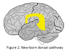
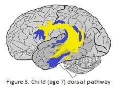
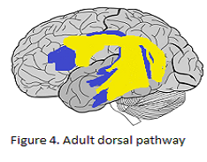
Bilingualism
Bilinguals v Monolinguals
Multilingual individuals must meet demands during language use that monolingual speakers are not faced with. Figuring out what language to speak and swapping between both of their languages requires additional structures.
Analysis on both surface area and thickness asymmetries were conducted. These showed that the anterior cingulate was the only region that differed reliably across groups. Bilinguals had significant rightward asymmetry compared to monolinguals who had more of a leftward asymmetry. These seem mainly due to variations in the thickness of the right hemisphere. Further tests which explored the effect of L1 proficiency, L2 proficiency, age and age of acquisition and their effects on anterior cingulate thickness asymmetry were run. L1 proficiency was shown to account for most of the variance, as bilinguals which greater L1 proficiency had less rightward asymmetry of the anterior cingulate. The bilinguals’ rightward asymmetry could be due to enhanced growth or reduced pruning of the right cortex in relation to left. This asymmetry could also reflect greater white matter expansion for the left cingulate in relation to the right. Recently it was discovered that the anterior cingulate is one of the cortical regions with a negative relation between cortical thickness and FA of the underlying superficial white matter. This finding indicates that thinner cortex in this area is associated with increased white matter integrity. However, thickness changes may be associated to both grey or white matter so further findings would be needed (Felton et al., 2017).
Experiments on the callosal volumes of monolinguals and bilinguals shows no significant difference in the total callosal volume but there are variances in the specific segments. Bilinguals are shown to have larger mid-anterior and central segments. Further analysis shows a significant correlation between the anterior cingulate asymmetry and the combined mid-anterior/central callosal volume. This provides more indication of altered interhemispheric organisation associated with language experience (Felton et al., 2017).
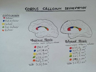
Figure 5. Corpus callosum segmentation monolingual female versus bilingual female
Early v Late Bilingualism
There are two types of bilingualism. Simultaneous bilinguals learn two languages at the same time from birth. Sequential bilinguals meanwhile learn their second language later on in life. People acquiring a second language at a later age are rarely able to obtain a native-like accent despite years of practice and high proficiency in other aspects of the language. Simultaneous bilinguals are usually able to speak both languages with a native-like accent although some may occur. For example, studies on Korean immigrants in America showed that children had less of an accent that the adults when speaking English. However, if they were over 6 years old they all still had some detectable accent after 4 years of immersion. 6-14 years old considered sequential bilinguals (Berken, J.A., et al., 2016).
Early bilingualism may lead to the development of new synapses, myelination and pruning of connections of neural circuits. The brain matures with a further development of neural networks. Neural maturation is especially extensive in the first few years of life so this is the period where it would be most sensitive to sensory experiences such as exposure to further languages and so would undergo the most neuroplasticity. When exposed to multiple languages from birth an infant has an increase in their complexity of sociolinguistic and sensorimotor processing so as to better interact with their environment. This leads to the optimal neuroplasticity period lasting longer. Simultaneous and sequential bilingual both undergo brain changes but the substrate it manifests on is different. Macroscopic differences in adults are subtle as they are more apparent on the microscopic level. Overall simultaneous bilinguals have enhanced cognitive and language processes which facilitate overall brain development. Most studies suggest optimal intervals to acquire languages, this being especially important for phonology which is the aspect most affected by age. However, others also argue that it is a progressive and linear decline of L2 proficiency potential with age.
The influences of bilingualism on tract specific mean fractional anisotropy (the property of being directionally dependent, value between 0 and 1 (0 means it is isotropic, so unrestricted or equally restricted in all directions), radial and mean diffusivities (the capacity measure of a substance or energy to pass through diffusion) was studied. The results showed that along the left direct segment of the arcuate fasciculus early bilinguals had significantly higher FA values while the late bilinguals had higher RD values. In the right posterior segment of the arcuate fasciculus late bilinguals had higher MD values than the early bilinguals. Further experiments on whether MD differences in the right posterior segment might be due to changes in fibre packing density were also carried out. Exploring MD and FA values for both groups showed a negative correlation for early bilinguals but not for late bilinguals. This suggests that the current effect most likely reflects increasingly dense and ordered packing of the fibre tracts in response to long-term early bilingual exposure the direct segment of the arcuate fasciculus supports phonological language functions hence changes in this segment could be due to early bilinguals facing increased phonological processing demands from an earlier age. Furthermore, the direct segment streamlines come from the inferior frontal gyrus, which is related to phonological language processing and is sensitive to grey matter density modulations related to bilingual exposure. Although some research proposes that white matter changes occur independently of grey matter changes, there seems to be a parallel between the present finding and the previous grey matter results. The most notable changes were observed along the left direct segment of the arcuate fasciculus. There early bilinguals exhibited significantly higher mean FA values than the late bilingual speakers. This finding helps link bilingual exposure to increased FA in several language-related white mater (Hämäläinen et al., 2017).
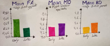
Figure 6. The average FA, MD and RD values from tracts
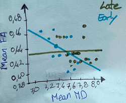
Figure 7. Correlation analysis between right posterior segment mean MD and FA values
Some studies have indicated that the right temporoparietal junction (which is the site of origin of the tracts of the posterior segment originate) might be important for higher-order cognitive processes involving both language and social cognition. Therefore, the right posterior segment may have some aspects of social functions. On this basis, it has been suggested that multilingual exposure facilitates effective communication skills as it was shown that bilingual children perform better than monolingual children on theory-of-mind tasks). It’s therefore likely that the higher fibre organisation found for early bilinguals along the right posterior segment could be due to enhanced socio-communicative aspects related to early bilingualism and multilingual environment (Hämäläinen et al., 2017).
Tract-specific TBSS analysis revealed a group difference bilaterally over frontal parts of the inferior fronto-occipital fasciculus, where early bilinguals had significantly higher than late bilinguals. This difference was the most pronounced over the right inferior fronto-occipital fasciculus. (Hämäläinen et al., 2017)
Furthermore laterality effects were also researched. The majority of bilinguals were shown to exhibit either an extreme left lateralisation or a strong leftward asymmetry of the direct segment of the arcuate fasciculus. In over half of the participants in the extreme left lateralisation group, the direct segment in the right hemisphere could not be traced consistently. Only a handful of participants presented a relatively symmetric left/right distribution of the direct segment (bilateral configuration). To evaluate the difference in the distribution of lateralisation pattern between the early and late bilingual groups a Fisher's exact test was performed an unequal distribution was revealed and further experiments showed that the extreme left lateralisation pattern was over-represented in late bilinguals. Furthermore, all the participants with a bilateral pattern belonged to the early bilingual group, hence an early multilingual environment contributes to a more bilaterally balanced structural configuration of the perisylvian language tracts. However other factors such as musical training and singing may also promote a larger and more structured right arcuate fasciculus composition (Hämäläinen et al., 2017).
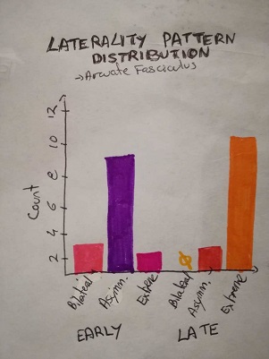
Figure 8. Laterality Pattern Distribution in the Arcuate fasciculus
Private Speech
Bilinguals’ private speech of seems to follow a similar developmental to that of monolinguals in the form of an inverted U shape curve relative to mental age. They also show greater use of task-relevant private speech on more difficult tasks and a path of gradual sub vocalisation and increasing internalisation. The use of private speech seems to be used the same for both monolinguals and bilinguals for reasoning, problem-solving and emotional expression. Social origins are a big factor influencing private speech for example the daily use of each language and proficiency. It’s usually influenced by the language previously used or if thinking of a concept then using the language in which this concept was first internalised (Sawyer, 2016).
However, some L2 proficiency, even at a bilingual level, might still mean a private speech in mostly one language. Usually in simultaneous bilinguals’ private speech is commonly in both languages but one becomes more dominant at preschool age depending on their social environment and proficiency. Only a very balanced bilingual has an equal use of both speeches, most bilinguals have a dominant tongue (Sawyer, 2016).
Code-switching (the ability to alternate two languages within the same conversation) can occur in private speech although it is quite rare and only in balanced bilinguals. Even if code-switching is seen as occurring rarely, it’s common for bilinguals to have a stream of private speech utterances in one language before switching to another stream in their second language (Sawyer, 2016).
Conclusion
As we can see, to this day, neural background in language learning is still a topic researchers have not been fully able to wrap their heads around despite making highly important breakthroughs that have been a gigantic leap forward in this area. We now know that the inferior frontal and temporal cortices are pivotal for language development but dominance can only be narrowed down to the left hemisphere. We also see that learning another language can be easy, as a child who is learning both languages at once, and much more difficult as an adult learning a second language later on in life. It is now known that the latter will find it harder to pick up the accent than one who has been learning the two languages from mother tongue. All in all, we can say that language learning is a diverse field in which researchers have quickly enhanced our knowledge on, however, we still have much more to unearth.
References
- Berken, Jonathan A., Vincent L. Gracco, and Denise Klein. "Early Bilingualism, Language Attainment, And Brain Development". Neuropsychologia 98 (2017): 220-227.
- Jens Brauer, Alfred Anwander, Daniela Perani, Angela D. Friederici. "Dorsal and Ventral Pathways in Language Development". Brain and Language 127.2 (2013): 289-295.
- Adam Felton, David Vazquez, Aurora I. Ramos-Numez, Maya R. Greene,Aleesandra Macbeth, Arturo E. Heranandez, Christine Chiarello. "Bilingualism Influences Structural Indices of Interhemispheric Organization". Journal of Neurolinguistics 42 (2017): 1-11.
- Friederici, Angela D. "The Cortical Language Circuit: From Auditory Perception to Sentence Comprehension". Trends in Cognitive Sciences 16.5 (2012): 262-268.
Sini Hämäläinen, Viljami Sairanen, Alina Leminen, Minna Lehtonen. "Bilingualism Modulates the White Matter Structure of Language-Related Pathways". NeuroImage 152 (2017): 249-257.
- Sawyer, Jeremy. "In What Language Do You Speak to Yourself? A Review of Private Speech and Bilingualism". Early Childhood Research Quarterly 36 (2016): 489-505.
Picture References
Figure 1. "Brodmanns's Area"- graphic from slide 287 of Neurophysiology lecture slides of Szent Istvan University Physiology department.
Figure 2. “The dorsal pathways of language network in new-born infants”- graphic from slide 173 of Neurophysiology lecture slides of Szent Istvan University Physiology department.
Figure 3. “The dorsal pathways of language network in children the age of 7.” - graphic from slide 173 of Neurophysiology lecture slides
Figure 4. “The dorsal pathways of language network of adults.” ”- graphic from slide 173 of Neurophysiology lecture slides
Figure 5. “The segmentation of the corpus callosum in a monolingual female versus a bilingual female”- based on (Felton et al., 2017)
Figure 6. “The average FA, MD and RD values from tracts.”- based on (Hämäläinen et al., 2017)
Figure 7. "Correlation analysis between right posterior segment mean MD and FA values"- based on (Hämäläinen et al., 2017)
Figure 8. "Laterality Pattern Distribution in the Arcuate fasciculus"- based on (Hämäläinen et al., 2017)
