|
Size: 16782
Comment:
|
← Revision 81 as of 2013-12-03 22:02:28 ⇥
Size: 16783
Comment:
|
| Deletions are marked like this. | Additions are marked like this. |
| Line 2: | Line 2: |
| '''The mechanisms of the MDMA''' |
|
| Line 4: | Line 6: |
| __'''The mechanisms of the MDMA'''__ |
The mechanisms of the MDMA
Contents
- Description
- Structure
- Effects
-
Mechanism
- MDMA as a substrate for monoamine transporters
- It exhibits the highest affinity for the Serotonin transporter SERT protein, and has less affinity for the dopamine (DAT) and noerpinephrine (NET) transporters
- MDMA stimulates the efflux of monoamine hormones
- MDMA inhibits tryptophan hydroxylase (TPH) the rate limiting enzyme for serotonin synthesis
- MDMA partially inhibits the Monoamine oxydase B
- MDMA Increases free cytoplasmic pool of serotonin
- Serotonin
- Dopamine
- Norepinephrine
- References
Description
MDMA - Ecstasy (Drug), Methylenedioxymethamphetamine. Derivates from the synthetic psychostimulant methamphetamine (METH).(Figure 1) It is a stimulant drug which induces hallucinogenic effects on users, with feeling of euphoria and a heightned perception. Its molecular aspect resembles the monoamine neurotransmitters Epinephrine(E) and Dopamine (DA). It mimetises the neurophysiological action of serotonin (5-HT), E and DA. (Holley et al, 2013)
Structure
Figure 1 - Structure of the MDMA above. MDMA is a member of the ampetamine family. Below, structure of other amphetamines, coming from the same family.(Hauw, 2013)
Effects
Immunosuppressive effect
Suppresses production of pro inflammatory cytokine tumour necrosis factor (TNF alpha), Interleukine (IL)-1beta, increases produc of the endogenous immunosuppressive cytokine (IL-10), promoting an immunosuppressive cytokine phenotype. MDMA also suppresses circulating Lymphocyte numbers, CD4+T cells being affected, alters T cell functions.(Connor, 2004)
Physchological and Physical effects
Sens of euphoria, wellbeing, sometimes Hallucination. Elevation in blood pressure, elevation of body temperature and enhanced tachycardia.
Mechanism
MDMA as a substrate for monoamine transporters
Inhibitor of SERT,DAT,NET MDMA affects both central and peripheral nervous system. It is said to be an indirect monoaminergic antagonist. Indeed, MDMA is a substrat for monoamine transporters and by this way, competes for the uptake of three neurohormones in the nerve terminal- Serotonin, Dopamine and Norepinephrine.
It exhibits the highest affinity for the Serotonin transporter SERT protein, and has less affinity for the dopamine (DAT) and noerpinephrine (NET) transporters
SERT’s activity is controlled by 3 different protein kinases - Protein Kinase C, Protein Kinase A, P 38 Mitogen activated protein kinase, controlling the phosphorylation state of SERT and its rate of insertion in the membrane.(Figure 2) The mechanisms underlying how MDMA directly affects the signalling networks responsible for SERT’s regulation are unclear. Studies have shown that MDMA could rapidly internalize SERT from the plasma membrane into the cystosol, decreasing SERT cell surface localization and function. It has been shown that the regulation by MDMA of the serotonin transporter SERT was done via the Protein Kinase C1.(Holley et al, 2013)
Figure 2 - Phosphorylation is a reversible PTM that regulates protein function. Left panel: Protein kinases mediate phosphorylation at serine, threonine and tyrosine side chains, and phosphatases reverse protein phosphorylation by hydrolyzing the phosphate group. Right panel: Phosphorylation causes conformational changes in proteins that either activate (top) or inactivate (bottom) protein function.(Hauw, 2013)
MDMA stimulates the efflux of monoamine hormones
Studies have found that MDMA directly and indirectly stimulates the efflux of the hormones for which it blocks the transporters of.(Rudnick and Wall, 1992)
1)MDMA stimulates the efflux of preloaded 5 HT serotonin,
2)To a lesser extent stimulates the efflux of dopamine DA
3)Interacts with monoamine transporters, stimulating non exocytotic release of 5HT DA and norepinephrine NE in rat brain. (Capela et al, 2009)
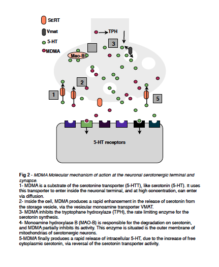
MDMA produces an acute and rapid enhancement in the release of 5 HT from the storage vesicles, by entering the vesicles via the monoamine transporter (VMAT) and reduces vesicular neurotransmitter stores via a carrier mediated exchange mechanism.
2 mechanisms of stimulation of the efflux of hormones
1)Transmitter molecules exit cell along their concentration gradients via reversal of normal 5 HTT function
Indeed, MDMA causes the transporters to work in reverse - MDMA is transported to the nerve cells, serotonin, dopamine and norepinephrine are dragged out.
2)Cytoplasmic concentration of transmitter are increased due to drug induced disruption of vesicular storage
This efflux is more complex: MDMA dissipates the interior acid in addition to its direct effect on the vesicular amine transporter. There is a PH difference generated by the vacuolar ATPase, which pumps H+ ions into the vesicle interior. Serotonin accumulation is driven by this pH difference, and dissipation of interior acid decreases serotonin accumulation and increases efflux.(Rudnick et Wall, 1992)
MDMA inhibits tryptophan hydroxylase (TPH) the rate limiting enzyme for serotonin synthesis
Tryptophan hydroxylase (TPH) is an enzyme (EC 1.14.16.4) involved in the synthesis of the neurotransmitter serotonin. TPH catalyzes the following chemical reaction L-tryptophan + tetrahydrobiopterin + O2 5-Hydroxytryptophan + dihydrobiopterin + H2O It is thought that this occurs because of oxidative stress which MDMA places on the neuron. This oxidative stress might occur through several possible channels. methylenedioxymethamphetamine, produced a rapid, persistent and dose-dependent reduction in cortical tryptophan hydroxylase activity, the rate limiting enzyme of 5-ht - irreversible inhibition. (Rattray, 1991)
MDMA partially inhibits the Monoamine oxydase B
Located in outer membrane of mitochondria of serotoninergic neurons - is the enzyme responsible for 5 HT degradation - its acitivity is partially inhibited by MDMA. Enzyme that catalyzes the following reaction : RCH2NH2 + H2O + O2 → RCHO + NH3 + H2O2. MAO, acting on primary, secondary and tertiary amines, and is involved in metabolism of important neurotransmitters - dopamine, serotonine, noradrenaline and adrenaline. Two isoforms exist in the brain MAO A (catecholaminergic neurons) and B (serotoninergic neurons, astrocytes and glia).
Oxydative stress related damage due to H2O2 will occur via the oxydative deamination of monoamine neurotransmitters by monoamine oxydase. H2O2 promotes an increase in hydroxyl radical HO formation, a higly toxic reactive oxygen species leading to the damage of cellular proteins, mitoochondrial DNA and proteins, which contributes ot MDMA neurotoxic events. After MDMA admin, increase of extravesicular levels of monoamine neurotransmitters inside nerve endings -MAO mediated metabolism of dopamine and serotonin leads to the generation of the reactive aldehyde intermediate 3,4-dihydroxyphenylacetaldehyde (DOPAL) and 5- hydroxyindole-3-acetaldehyde (5-HIAL), respectively, before their conversion into the more stable metabolites 3,4-dihydroxyphenyl- acetic acid (DOPAC) and hydroxyindole-3-acetic acid (5-HIAA) by aldehyde dehydrogenase (ALDH). (Capela et al, 2009)
MDMA Increases free cytoplasmic pool of serotonin
MDMA promotes a rapid release of intracellular 5 HT to the neuronal synapse, via reversal of the 5 HTT activity. Due to this the level of serotonin dramatically increases.(Figure 3) Nerve cells become overstimulated. (affecting mood sleep perception appetite).

Figure 3 - Mechanisms of action of the MDMA.(Hauw, 2013)
Serotonin
There is a general agreement that MDMA produces a substained long term neurotoxic loss of 5-HT nerve terminals in several brain regions of rats, guinea pigs and several species of non human primates. On the other side, the same dose that produces a major loss of 5-HT produces no long term neurotoxic loss of cerebral dopamine content in these species. In contrast- MDMA is generally accepted to behave as a selective dopaminergic neurotoxin in mice in the striatum. (Colado et al, 2001)
Dopamine
It has previously been excepted that MDMA affects the extracellular dopamine and serotonin levels in the brain thus resulting in many of its psychological symptoms or often disastrous consequences. Hagino, Takamatsu et al were able to experimentally deduce from mice experiments that dopamine and serotonin transporters are the main site of action for this psychedelic amphetamine. Knockout mice were used as test subjects and it was shown that upon administration of MDMA normal mice expressed an increased in dopamine and serotonin levels in the striatum and prefrontal cortex of their brain. This increase of extracellular hormones was not registered or was in such a negligible amount within the knockout mice thus enabling conclusion that MDMA acts both at dopamine and serotonin transporters(Hagino et al, 2001). A further study focused on activity, substrate binding and cell surface expression of the dopamine reuptake transporter (DAT) on the pre-synaptic neurone membrane. In this study that focused on cocaine and amphetamines as effective drugs, this family of drugs could be said to encompass the psychedelic amphetamine MDMA. In a normal environment, before the administration of any inhibitors or drug, DAT is expressed on both the cell surface and the intracellular membranes of brain cells. It was shown that upon administration of amphetamines the drug would compete with dopamine for binding with the transporter thus reducing the level of dopamine which can enter the cell, another method of the reduction of intracellular dopamine is the amphetamine-induced down regulation of DAT on the cell membrane. Both of these mechanisms result in reduced movement of dopamine into the cell thus resulting in an extracellular build up on dopamine.(Kahlig et Galli, 2003).
Rats post exposure to high doses of dopamine (approx 10mg/kgbw x4 doses) was also studied. Here they concluded that although dopamine levels themselves are not significantly altered compared to levels in rats subjected to a low dose of MDMA, the presence of dopamine metabolites is dramatically reduced (DOPAC 34.3%, HVA 33.5%). As a result it can be noted that the dopamine turnover is effected by the exposure to high doses or MDMA, this may lead to a disastrous oxidative stress. However you could ask if these effects are permanent, it was also shown that even after removal of all amphetamine drugs by a washout the extracellular dopamine levels remained high, this can be attributed to the low number of dopamine transporters on the cell surface. (Figure 4) It seems that this lack of transporter expression is a long-lasting effect of drug use which may be account for why recovery after drug use is so prolonged (this recovery will be discussed later). A third mechanism of amphetamine upon the pre-synaptic membrane is the binding to amphetamine transporters in the membrane as endocytosis into the neurone. Once inside the neurone amphetamines stimulate the exocytosis of dopamine from vesicles into the synaptic cleft, this adds to the increase of dopamine concentration in the cleft. Studies have shown the effect of this released dopamine may be hyperthermic or hypothermic. (Biezonski et al, 2013)
Norepinephrine
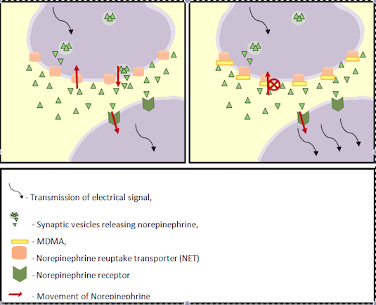
Moron et al (2002) also found a correlation between the presence of NET (norepinephrine reuptake transporter) inhibitors and increased extracellular dopamine in the prefrontal cortex. It was then postulated that MDMA may act at NET and increase extracellular dopamine concentrations. NET is a transporter in the pre-synaptic membrane responsible for the sodium- chloride dependent reuptake of extracellular norepinephrine aswell as having a side effect on the reuptake of dopamine. The reuptake of these two hormones from the synaptic cleft is key for normal functioning of a synapse and hence central nervous system activity. This monoamine transporter is often the target for antidepressant drugs enabling serotonin and other mood improving hormones to remain at a high extracellular level, this affect is mimicked when taking recreational drugs. The transport of epinephrine from the synaptic cleft into the cell only occurs upon binding of sodium and chloride ions acting as a ligand-gated ion channel. In order for the movement of ions out of the cell a concentration gradient must be generated with the aid of the sodium/potassium/ATPase pump. It has been shown that MDMA may lead to a reversal in the action of NET leading to a high extracellular norepinephrine concentration. A study was more able to specifically pinpoint the norepinephrine transporter effected by MDMA to the following 3 receptor types. (Table 1)
Receptor |
Effects |
central alpha(2A)-adrenoreceptors |
vasoconstriction to reduce heat loss |
peripheral alpha(1)-adrenoreceptors |
vasoconstriction to reduce heat loss |
beta (3)- adrenoreceptors in Brown Adipose Tissue |
Increase heat generation |
Table 1- Effects of 3 diffferent receptors on the heat system. (Docherty et Green, 2010)

Figure 4 - Mechanism of action of the amphetamine (MDMA) on two neurohormones - dopamine and noradreanaline (Catherine Hauw, 2013)
References
Figures
1- Hauw, 2013 - Figures 1,2,3,4.
