Effects of psychosine on the vascular system
Abstract
Psychosine is a cytotoxic glycosphingolipid which has both metabotoxic and cytotoxic effects on the living cells. Accumulation of psychosine is happening mainly in the nervous- and vascular system, which leads to harmful effects on the individ. Psychosine has major importance in diseases such as globoid cell leukodystrophy (krabbe disease) and in cell membrane architecture alteration. Different experiments has been done to gather more information about the influence of Psychosine in the different cells.
Introduction
|
Figure 1 Psychosine |
Psychosine is a subtype of glycolipids containing the amino alcohol sphingosine and is considered a sphingolipid because of the attached carbohydrate (Figure 1). Sphingolipids are compounds that play an important role in cell recognition and signal transduction. Sphingolipidoses is the name of disturbances in the sphingolipid metabolism, which mainly has an impact in the neural cells and tissues. It is related to lipid storage disorders, which we among other diseases can find Krabbe’s disease. Psychosine accumulated in the brain of humans, dogs or mice are affected (Igisu, 1989).
General
Sphingolipids is most often known for being part of the protecting plasma membrane lipid bilayer, which serves as a protection of the cell from harmful environmental factors. Glycosphingolipids, which psychosine is a subtype of, has an important role in the recognition and signaling of cells. Cell recognition is purely based on the sphingolipid properties and characteristics, whereas the cell signaling is based up on the interaction between the neighboring cells or with proteins. Because of its toxicity to living cells, psychosine is also referred to as a cytotoxic sphingolipid. It reveals its effect when it accumulates in the globoid cell leukodystrophy and alters the membrane architectures (Hawkins-Salsbury et al, 2013).
Psychosine as a toxic molecule
Psychosine is a part of the biosynthesis of cerebrosides and the compound is an intermediate in the reaction between; sphingosine, UDP-galactose and fatty acid- coenzyme A. psychosine is a very cytotoxic lipid, which is capable of inducing cell death in a broad variety of cell types. One type of cell which is the most commonly affected cell by this lipid is; Oligodendrocytes (Figure 2) which is related to the Globoid Cell Leukodystrophy (GLD) (Won et al, 2013). Psychosine induces pleiotropic effects, which means that it may have an effect on several different genetic traits. It can lead to dysfunction in several different cellular pathways and compartments with diffuse connections between them. The accumulation of psychosine in the nervous system is especially affecting the oligodendrocytes, which leads to continuous increasing demyelination and infiltration of macrophages/monocytes into the central nervous system. This is what happens in Krabbe’s disease. The accumulation of psychosine in the cell membrane alters the membrane architecture, this is possible because of its affinity to cholesterol. Cholesterol is a natural part of the cell membrane which keeps the phospholipids from getting too close and gelling together (Hawkins-Salsbury et al, 2013).
Psychosine can both purpose its effects as a neurotoxin or as a metabotoxin. As a neurotoxin, psychosine disturb or attack tissue or cells in the nerve system. Also, as a metabotoxin, psychosine is a metabolite derived internally that causes unfavorable health effects on chronically high levels. An example of psychosine working both as a neurotoxin and a metabotoxin is the neurological disease globoid cell leukodystrophy (GLD) also known as Krabbe disease. Psychosine accumulates in the oligodendrocytes and causes disruption on the nervous system. This happens because the lysosomal enzyme responsible for removing psychosine, galactosylceramidase (GALC) is absent. This will lead to chronically high levels of psychosine, and globoid cell leukodystrophy develops (GLD) (Won et al, 2013).
|
Figure 2 Oligodendrocyte |
Experiment of psychosine on schwann cells
An experiment was performed by checking the effects of psychosine on the schwann cells. This was carried out by studying cultures of schwann cells in a medium containing different concentrations of psychosine. "1, 10, 50, 75 and 100 microM psychosine for 24, 48 and 72 hours (h). When incubated in 50-100 microM concentrations of psychosine for 24 h, 52-99% of cultured Schwann cells did not survive. Lower concentrations (1-10 microM) did not significantly reduce Schwann cell numbers for the first 24 h. However, only 43-69% of cultured Schwann cells survived in these low concentrations for 48 h, and substantially fewer remained after 72 h of incubation." This experiment was performed by the Laboratory of Experimental of Neuropathology, National Institute of Neurological Disorders and Stroke, National Institutes of Health, Bethesda, Maryland 20892 (Tanaka and Webster, 1993).
After the experiment the schwann cells were examined. Cytoplasm of the schwann cells exposed to psychosine showed several membranous inclusions and fewer mitochondrias, where the ones left showed swelling. The findings in the experiment did support the hypothesis that psychosine accumulated in the globoid cell leukodystrophy is toxic for schwann cells, and alters their capacity to maintain the fatty insulation, myelin (Tanaka and Webster, 1993).
Krabbe disease
|
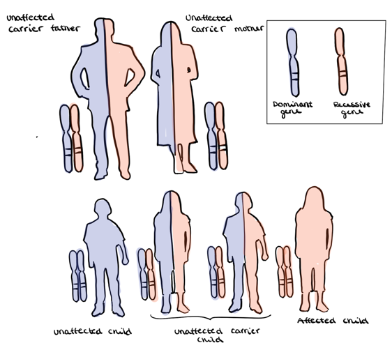
Figure 3 Autosomal recessive inheritance |
Krabbe disease (KD), or globoid cell leukodystrophy, is a rare neurodegenerative disorder which is caused by galactocerebrosidase (GALC) enzyme deficiency. The GALC is an enzyme essential for myelin turnover. It is an autosomal recessive disease, which means that both of the parents must have been carriers of the disease. The incidence of the disease in the United Stated is about 1 in 100 000 individuals (Escolar et al, 2017; Pellegrini et al, 2019).
Signs and symptoms
In most cases the signs and symptoms of Krabbe disease will develop in within the first 12 months of life, which is called early-infantile Krabbe disease (EIKD). This form is characterized by excessive crying, irritability, stiffness, and developmental delay before the age of 12 months. The neurodegeneration will occur rapidly in infants with this type. Untreated infants usually die during early childhood. To a lesser extent, the cases happens after the first year of life, which is called late infantile Krabbe disease (LIKD). This form is characterized by slow development, vision loss, slurred speech and gait abnormalities. Late infantile, late juvenile and adult form is characterized by milder progression and severity, but is highly variable (Escolar et al, 2017; Orsini et al, 2000).
Krabbe disease is a neurodegenerative disorder, leading to a disruption of myelin sheath in the central nervous system (CNS) and peripheral nervous system (PNS). Myelin is important to protect the nerve cell. The myelin sheath works as a plasma membrane around the nerve axon and ensures rapid transmission of nerve signals in the brain and throughout the nervous system. Without this protection the brain cells will die and other parts of the body will not work properly (Escolar et al, 2017; Morell and Quarles, 1999; Pellegrini et al, 2019).
Causes
The disease occurs due to mutations in the galactocerebrosidase gene. These mutations lead to a reduced activity of the enzyme; galactocerebrosidase (GALC). This enzyme is necessarily to break down toxic substances in nerve tissues. Defective GALC will lead to weakened degradation of the two glycolipids: galactosylceramide and psychosine (galactosylsphingosine). The build-up of psychosine in the myelin sheath of the nervous system can be a biomarker to detect Krabbe disease. The accumulation of psychosine in the nervous system and widespread degeneration of oligodendrocytes and Schwann cells, will cause rapid demyelination. Demyelination will cause inhibition of nerve impulses, so the brain cannot send nerve impulses to the rest of the body. This will result in disability (Escolar et al, 2017; Morell and Quarles, 1999; Pellegrini et al, 2019).
As the psychosine continues to builds up in the nerve tissues and destroy the myelin – the disability will continue to increase and the brain will lose control over the body. Representative cases are movement, vision, speech and life essential functions like maintaining heartrate and respiration. Eventually it will cause death (Escolar et al, 2017; Pellegrini et al, 2017).
Treatment
Currently, there is no long term treatment or definite cure for the infants that has already developed symptoms of Krabbe disease. Therefore, the only treatment available is the hematopoietic stem cell transplantation (HSCT). It has shown to slow down the development of the disease, and to preserve cognitive skills of patients who developed the disease at early-infantile state (EIKD). HSCT will only be effective if the patient does not have any neurological damage prior to the transplantation. It does not have a reversing effect if the damage is already done, survival may be extended, but most patients will continue to deteriorate. Other treatments for Krabbe disease are under investigation in animal models. Including gene therapy, substrate reduction therapy, chemical chaperones and enzyme replacement therapy. However, a population-based newborn screening (NBS) for the disease is probably the most efficient way to detect the disease before symptoms are shown and the nerve tissue is damaged (Escolar et al, 2017; Pellegrini et al, 2019).
Psychosine in the vascular system
The microvascular endothelium of patients with globoid cell leukodystrophy and twitcher mice, has shown alterations in function and architecture due to the accumulation of psychosine. Twitcher mice is widely used in the investigation of Krabbe disease and other diseases, due to its similarities with human beings. Krabbe disease, in addition to neural damages shows alterations in the vascular system (Cappello et al, 2016).
Experiment – Psychosine’s effect on endothelial cells in twitcher mice
An experiment was performed on twitcher mice and wild type mice, where the lower motor system was investigated. The experiment was performed before onset and after complete development of the disease. Endothelial cells of the blood vessels in twitcher mice showed alterations compared to the wild type mice upon complete development of Krabbe disease. In physiological cases, the basal membrane should be continuous, with tight junctions and surrounding pericyte processes. In contrast, the endothelial cells of twitcher mice contained certain regions with thinner basal membrane, and some protrusions into the lumen of the endothelial cells. Based on this experiment, the twitcher mice showed pathological signs in contrast to the wild type mice (Capello et al, 2016; Belleri and Presta, 2016).
Angiogenesis
Psychosine is a molecule having an effect on the angiogenesis, where alterations in the migration of endothelial cells and the invasion of the extracellular matrix are some representatives (Belleri et al, 2013). Since the migration of endothelial cells are crucial for angiogenesis, psychosine is thereby able to inhibit this process (Belleri et al, 2013). A wound healing assay containing murine aortic endothelial cells was investigated, where the purpose was to analyze the migration of cells in a monolayer cell culture (Jonkman et al, 2014). Psychosine containing endothelial cell monolayers showed non-proper migration of the cells (Belleri et al, 2013). Normally after migration and mechanical wounding, the cell microtubule organization center has changed its location from a random one to ahead of the nucleus (Belleri et al, 2013). This new location should be in the direction of the migration. Monolayers containing psychosine showed significantly slower migration than cells under physiological conditions, like those without the cytotoxic molecule. In the course of psychosine accumulation, the molecule is able to alter the CNS vascularization as well (Belleri et al, 2013). Based on this information, psychosine exerts its effects both in the angiogenesis and in the CNS vascularization, which is partly responsible for the Krabbe disease (Belleri et al, 2013).
Proliferation of endothelial cells
Psychosine has an inhibitory effect on the proliferation of endothelial cells. This phenomenon was proven in an experiment with murine aortic endothelial cells. Three experiments were performed, one short term for 24 hours, and two long term proliferation assays for 72 and 120 hours. In the short term assay the ID50 was equal to 60 uM. Contrary, the long term assays containing ID50, turned out to be 15 uM and 5 uM. This experiment shows that psychosine is more effective over time. The negative control was N-acetyl-D-sphingosine, which did not prevent proliferation like psychosine. Thereby, the conclusion is that accumulation of psychosine in the organism due to GALC deficiency, has an inhibitory effect on the proliferation of endothelial cells (Belleri et al, 2013).
VEGF and FGF2
Neovascularization is the formation of new blood vessels, which is very important for the central nervous system (CNS), including the development and protection. Vascular endothelial growth factors (VEGF) and fibroblast growth factor 2 (FGF2) are important angiogenic factors, both for the neural cells and the endothelial cells of the CNS. They are major substances in the production and protection of neurons, and also in angiogenesis. FGF2 can be used to induce angiogenesis. In an experiment concerning the effect of psychosine in the neovascularization in vivo, FGF2 was absorbed with psychosine in gelatin sponges, and inserted in an embryo on top of the chorioallantoic membrane. The result showed significant inhibition of FGF2 on the neovascularization in the presence of psychosine. In contrast to the control samples which only contained the FGF2 and the gelatin sponges, showed normal function of FGF2 on the neovascularization. Based on this, we can conclude that psychosine has an anti-angiogenic property, which in other words inhibits the angiogenesis (Belleri et al, 2013).
Endothelium of infected twitcher mice, showed in an experiment reduced ability to respond to vascular endothelial growth factor (VEGF). This phenomenon was due to psychosine accumulation, which inactivates the mitogenic and motogenic response of the endothelial cells. In all, this is very unfavorable because the vascular endothelial growth factors are important signaling proteins in both vascular and neuronal functions. Representative cases are during embryonic development, in the formation of new vessels after injury and in neuronal proliferation. With the decreased response of the endothelial cells to the VEGF, non-physiological alterations can occur in the endothelium of infected individuals (Belleri et al, 2013).
Conclusion
Psychosine is a glycosphingolipid which has important roles in signaling and recognition of cells. The compound is known to be cytotoxic to living cells, and expresses both its neurotoxic and metabotoxic effects in Krabbe disease. This disease is also known as globoid cell leukodystrophy (GLD), which occurs due to the lack of galactosylceramidase (GALC). This phenomenon results in accumulation of psychosine. An experiment was performed with different concentrations of psychosine on schwann cells. The result showed decreased capacity to maintain myelin in psychosine containing cells. Krabbe disease is a lethal disease with neural clinical signs, to which no effective treatments is developed yet.
Accumulation of psychosine also has its effect on the vascular system, by having an anti-angiogenic effect. This phenomenon is due to defects regarding fibroblast growth factor 2 and vascular endothelial growth factors. In addition to this, psychosine can have a negative effect on the angiogenesis by significantly decrease the migration of endothelial cells.
By this research, there is reasons to believe that psychosine exerts its negative effects both in the nervous system and in the vascular system.
References
Belleri, M.; Presta, M. (2016): Endothelial cell dysfunction in globoid cell leukodystrophy. Journal of Neuroscience Research 94: (11) 1359-1367
Belleri, M.; Ronca, R.; Coltrini, D.; Nico, B.; Ribatti, D.; Poliani, P. L.; Giacomini, A.; Alessi, P.; Marchesini, S.; Santos, M. B.; Bongarzone, E. R.; Presta, M. (2013) Inhibition of angiogenesis by ß-galactosylceramidase deficiency in globoid cell leukodystrophy. Brain 136: (9) 2859-2875
Cappello, V.; Marchetti, L.; Parlanti, P.; Landi, S.; Tonazzini, I.; Cecchini, M.; Piazza, V.; Gemmi, M. (2016): Ultrastructural Characterization of the Lower Motor System in a Mouse Model of Krabbe Disease. Scientific reports 6: (1) 1-13
Escolar, M.; Kiely, B.; Shawgo, E.; Hong, X.; Gelb, M.; Orsini, J.; Matern, D. and Poe, M. (2017): Psychosine, a marker of Krabbe phenotype and treatment effect 121: (3) 271-278
Hawkins-Salsbury, J.A.; Parameswar, A. J.; Jiang, Xuntian.; Schlesinger, P. H.; Bongarzone, Ernesto.; Ory, D.S.; Demchenko, A. V.; and Sands, M. S. (2013): Psychosine, the cytotoxic sphingolipid that accumulates in globoid cell leukodystrophy, alters membrane architecture. J Lipid Res 54: (12) 3303-3311
Igisu, H. (1989) : Psychosine: a “toxin” produced in the brain –its mechanism of action. J UOEH 11: (4) 487-93
Morell P, Quarles RH. (1999) The Myelin Sheath. In: Basic Neurochemistry: Molecular, Cellular and Medical Aspects. 6th edition. Philadelphia: Lippincott-Raven ISBN-10: 0-397-51820-X
Jonkman, J. E.; Cathcart, J. A.; Xu, F.; Bartolini, M. E.; Amon, J. E.; Stevens, K. M.; Colarusso, P. (2014) An introduction to the wound healing assay using live-cell microscopy. Cell Adh Migr 8: (5) 440-451
Orsini JJ, Escolar ML, Wasserstein MP, Caggana M (2000) Krabbe Disease. In: GeneReviews® Seattle (WA): University of Washington, Seattle ISSN: 2372-0697 (accessed on 2019.05.19.)
Pellegrini, D.; Del Grosso, A.; Angella, L.; Giordano, N.; Dilillo, M.; Tonazzini, I.; Caleo, M.; Cecchini, M.; McDonnell, L. (2019): Quantitative Microproteomics Based Characterization of the Central and Peripheral Nervous System of a Mouse Model of Krabbe Disease 18: (5) 1-57
Tanaka, K., Webster H. D. (1993): Effects of psychosine (galactosylsphingosine) on the survival and the fine structure of cultured Schwann cell 52 (5): 490-498
Won, JS.; Kim, Jinsu.; Paintlia, M. K.; Singh, Inderjit.; and Singh, A. K. (2013): Role of endogenous Psychosine accumulation in oligodendrocytes Differentiation and survival: Implication for krabbe disease. Brain Res 1508: 44-52
![]() End of edit conflict
End of edit conflict


