Elevated muscles enzymes in racehorses
Introduction
Thoroughbred racehorses perform intensive work each day in order to meet the high performance requirements that are expected by them to perform on the racetrack.
These horses are typically aged between two to five years old, though some can race for more prolonged periods.
Various changes in physiological parameters are associated with overtraining, which can be a serious problem for human and equine athletes (Hamlin M. 2010). This report is to investigate the elevated muscle enzymes on Racehorses in training.
|
Enzymes that are studied are aspartate aminotransferase (AST) and creatine kinase (CK). We will discuss muscle physiology, the mode of action, the clinical signs of lameness and other issues such as ‘tying up’ that results due to the muscle damage caused by these enzymes. Finally we will discuss what happens when elevated levels in the blood serum and the best treatment protocol to follow when these issues arise.
Molecular aspect of muscle Physiology and the coupling of contraction
Giving a small overview on the molecular aspects of muscle contraction will help us understand the mechanism better.
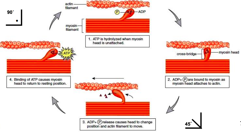
On the surface, a tropomyosin molecule covers the active state which could stimulate ATPase troponin complex which once activated with Ca2+, will displace the tropomyosin and slide into the groove of 2 actin molecules initiating the cross-bridge cycle.
Once the cross-bridge cycle starts, the actin-myosin complex is formed and will split the bound ATP which will cause an energy release.
This energy release causes the myosin head to move, and two power strokes will happen:
1. Power stroke 1: Myosin head tilts 40o but it is still glued to actin.
2. Power stroke 2: ATP dissociates further and a 5o conformational change happens.
However, this is all under the control of ATP and Ca2+ so any deficiency in one of them will lead to problems.
- In Ca2+ deficiency, no binding will happen and will therefore cause Parturient paresis
- In ATP deficiency, no dissociation can occur, and it will lead to rigor mortis.
|
Figure 2 – Cross bridge cycle in muscle physiology
Biochemistry background reactions
Tying-up is simply muscle cramps, the back and hindquarter muscles are most often affected by a combination of different mechanisms, leading to the accumulation of lack of muscle oxygenation, lactic acid and even the death of muscle cells.
This can be triggered by a strenuous exercise in an unfit horse, stress, and a dietary imbalance such as a diet that is high in cereals containing a high ratio of potassium and sodium, or on the other hand a diet that is deficient in various minerals and vitamins.
It is easier to prevent rather than to cure, therefore a diet of high fats and low carbohydrates is used. There is speculation that this may be a genetic trait due to some blood lines producing offspring with this tying up. (Valberg, MacLeay and Mickelson, 2022).
The greatest amount of AST is found in the liver, heart muscle and skeletal muscle and then small amounts found in the pancreas, erythrocytes and the kidneys.
AST catalyses the reaction between aspartate amino acids and glutamate and plays a role in the amino acid metabolism as previously taught in biochemistry.
|
Figure 3 – AST reaction
As AST is found in most cells in the body and when its concentration is increased in the serum, we assume this is due to damage of the liver or muscle due to leakage especially after intensive work out. However it is also able to increase in the serum due to hemolysis of the erythrocytes. (SKENDERI et al., 2022)
Aspartate aminotransferases (AST) is a group of enzymes that can catalyse the conversion of amino acids by transferring of the amino groups. Aspartate aminotransferase (AST), previously known as glutamate oxaloacetate transaminase (GOT), are the two most common aminotransferases of significance. Pyridoxal -5- Phosphate functions as the coenzyme in the amino transferring reactions. All amino transfer reactions contain, 2-oxoglutarate and 1-glutamate serve as one amino group acting as both acceptor and donor pair. (Rudat, Brucher and Syldatk, 2012)
Creatine kinase (CK) also increases due to liver or muscle damage therefore both of these (AST and CK) can be used to allow us to identify the exact cause of AST increase. Whereby both are increased together, it demonstrates muscle damage, but if AST is increased but the CK level stays the same this tells us we have liver damage. It takes the AST level to decrease very slowly after a muscle injury therefore we cannot know for sure if a high level is from a previous muscle injury or a present one. (Gwaltney-Brant, 2016)
|
Figure 4 – Creatine kinase reaction
Syndromes and diseases
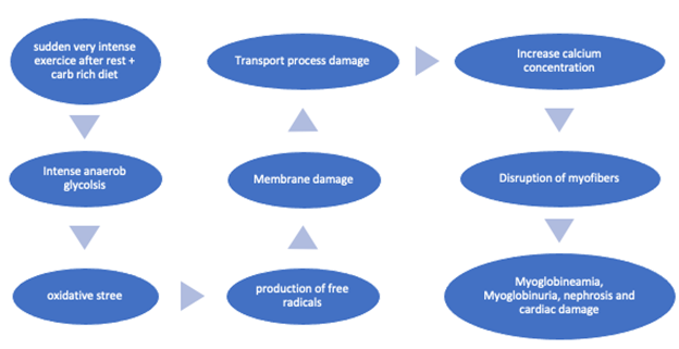
The most important muscular disorder is rhabdomyolysis, also called monday morning disease and equine paralytic myoglobinuria. This disease can be life- threatening as it causes muscle breakdown and muscle death. It is a serious condition that can be fatal or result in permanent disability which is a great problem because most horses are used in races and as a way to make money which is unfortunate for the horse seeing as he won’t be useful for the owner anymore.
The course of the disease is as follows:
|
Figure 5 – Rhabdomyolysis disease
Some diagnostics tests that are done in order to determine the disease are:
1. Blood tests:
· Measuring the level of substances that are released after damaged muscles.
· Three muscles that are always measured are CK, AST and LDH
1. Urinalysis
· Test the urine for muscle breakdown
· If positive, myoglobin which transports oxygen is released
1. Exercice tests
· Blood sampling after a period of controlled exercise
· Mild increase is normal, but after a certain level it is most probably a muscle disease
1. Muscle biopsy
· The myoglobin fibers would be damaged therefore a biopsy can be taken in order to find out.
Clinical signs
There has been research done into the elevated high levels of aspartate aminotransferase (AST) and creatine kinase (CK) in blood which has generally been considered to be an indirect marker of muscle damage (Baird .M 2012).
The elevated levels that were discussed earlier were shown to have a significant correlation with high CK (>100 iu/litre) and AST (> 300 iu/litre). Thoroughbreds in training regularly get blood samples taken to evaluate plasma (AST/CK) activities. Interestingly, fillies are more likely to have elevated CK and AST than colts. It is commonly observed that tying up occurs more frequently in young fillies than in colts or geldings, especially nervous fillies that are fed high grain diets such as oats for example.
If such fillies have been transported for many hours, prior to racing they can essentially “compete before the race” and experience tie-up. Also horses in poor condition are more susceptible to this, if they become dehydrated and experience a deficiency in potassium, magnesium, selenium or sodium. (Beech, 1997)
- The results show no relationship between elevations in serum muscle levels at the same time as the oestrous cycle, which would suggest that hormones that are involved in the cyclic cycle have no effect on the condition (Frauenfelder H.C 1986).
It is also interesting that in the horses tested, the two-year-olds tended to have higher AST activities than three-year-olds. Time of year had no significant effect on the number of animals with high or low activities (PAT. A 1990).
The rest and exercise relationship is interesting, as horses that were tested after a lower intensive day tended to have higher CK on a Monday after a weekend of rest, which could be due to the changes in the membrane permeability of skeletal muscle cells (Pickar J.G 1991). This would clarify the common name chosen for rhabdomyolysis as “Monday morning disease”.
These clinical signs of muscle stiffness and pain were observed in horses and could have been misinterpreted as lameness, the horses that showed signs of rhabdomyolysis are stiff all over and appeared to be ‘Jarred up’ which is a state where the horse is finding it difficult to move in a fluent manner. As opposed to lameness when there is a clear sign that there is injury to a specific limb instead of systemic muscle damage that can be experienced in multiple areas.
When biopsies were examined, fibres on an electron microscope contained disrupted myofibrils and swollen mitochondria.
This could suggest that horses clinically diagnosed with lameness and an increase in muscle enzymes is the main cause of repeated subclinical episodes of rhabdomyolysis after exercise.
Rather than leakage due to abnormal sarcolemmal permeability. (Valberg S 1993) Rhabdomyolysis is one of the main diagnoses of elevated AST and CK levels along with many other diseases.
Treatment plan
To treat tying-up in horses, managing the diet is a big part of it.
- Feeding a diet that provides 40% of DE in the form of starch may lead to more exercise-induced muscle damage than when lower-starch diets are fed
- Replacing starch with a specifically designed fat rations allows horses to continue a high-calorie intake with a decreased chance of muscle damage.
- Heart rate may give an indication of an animal’s nervous reaction to his surroundings and nervous horses are more prone to tying up. In a study, a fat diet was associated with lower heart rates and calmer behavior which suggests that replacing dietary starch with fat may decrease the incidence.
- Another study showed that high CK levels when they were exercised on Monday following two idle days increased the risks
=> Therefore, we can say that to have a well-managed horse with decreasing risks of rhabdomyolysis may require a high-fat, low-starch diet with a good exercise plan which will not allow excessive periods of rest and confinement (Equinews)
Other methods of treating this disease is:
- Give additional electrolyte, trace minerals and muscle buffers. Firstly, to replace electrolyte loss through sweating by giving one tablespoon of Epsom salts in the meal in the morning and night.
- Secondly it can be advised to supplement the feed with 50 to 60 grams of chromium and selenium and even adding vitamin E.
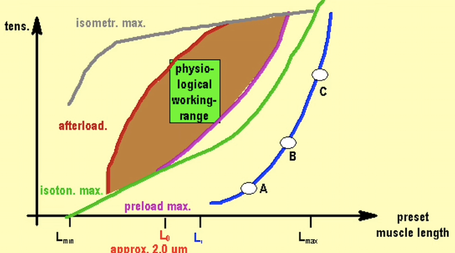
- Despite these deficiencies that can lead to tying up it can significantly reduce muscle strength.
|
Figure 6 – Working range of muscle
- Lastly (muscle buffers) supplementing roughly 50 millimetres in through the feed daily, an example of this is a solution of Neutradex which contains an acid buffer that ultimately neutralises the lactic acid that is produced during intense training or exercise.
Field research
When training racehorses, you have to treat all the horses as individuals. They all experience different effects of the high intensity of galloping everyday ‘routine work’. This starts at an age as young as two years old when a horse is ‘backed’ (saddle is put on a horse for the first time).
You can notice clinical signs as early as the first day that they do a stronger gallop after light canters. This happens as the muscles are put under pressure at a very young age.
Coming from a horse training background it is regular that blood tests can be carried out for many reasons such as general health, illness or fitness and it is done right before race day.
Out of our own interest and for the purpose of this study, we decided to carry out our own investigation. The horse tested in question called “Mariestella” had been showing clinical signs of Rhabdomyolysis.
Mariaestelle was a 3 year old thoroughbred filly that has been showing stiffness. The stiffness was first seen after her first bit of intensive work and then it was seen after routine work and worse again after a day of rest.
This now can be explained due to research carried out by Pickar as discussed earlier (Pickar J.G 1991) where he stated that this is due to the changes in the membrane permeability of skeletal muscle cells and once the muscle is injured it is only going to show worse after incorrect treatment and continuing galloping even if it is just routine work.
|
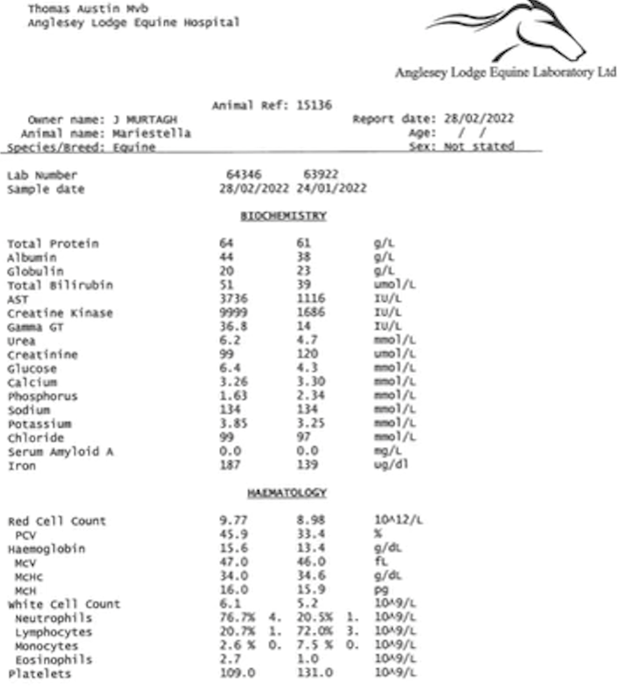
Figure 7 – Blood test of “Mariaestella”
As you can see from the blood test taken, the filly showed elevated AST and CK levels of :
- CK - 9999 IU/L
- AST - 3736 IU/L
The results explained a lot due to her performance at home and on the track. Even though she was not showing clinical signs, the injury was still there when she was running, her track performance was poor. Therefore even if a horse is not showing clinical signs, the previous injury can still be affecting performance.
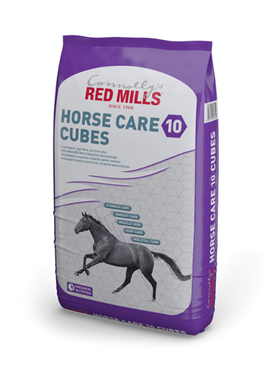
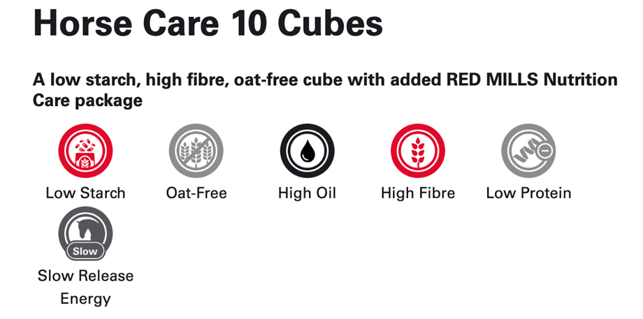
After these results were analyzed the diet of the filly was changed to 10% Horse Care, Red mills horse cubes. The ingredients include : Soya (Bean) Hulls, (Sugar) Beet Pulp, Barley, Wheatfeed, Alfalfa Meal, (Sugar) Cane Molasses, Maize, Soya Bean Extruded, Soya Oil, Sunflower Seed Meal, Mono-dicalcium Phosphate, Maerl 0.75%, Sodium Chloride, Magnesium Oxide, Mannan-/Fructo-oligosaccharides 0.1%.
|
Figure 8 – Red mills food bag
|
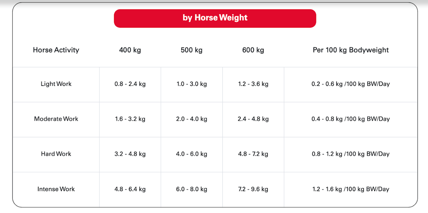
The feeding guidelines for horse weight according to Red Mills, taking into account the intensity of the training. Horses are fed little and often 4 times a day in our yard.
|
Figure 9 – Nutritional value of the feed
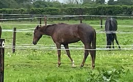
The weather also improved and this meant that after routine work she was put out in the field. We can see the nervy filly here laid back
|
Figure 10 - Mariaestella (owned photo) after healing
Summary
The elevated muscle enzymes that are studied are aspartate aminotransferase (AST) and creatine kinase (CK), which have a range of consequences.
Tying-up can also be simply called muscle cramps, of the back and hindquarter muscles are most often affected by a range of different mechanisms, resulting in the accumulation of lack of muscle oxygenation, lactic acid and even the death of muscle cells.
Aspartate aminotransferases is a group of enzymes that catalyse the conversion of amino acids by transferring of the amino groups. AST that was previously known as glutamate oxaloacetate transaminase (GOT), are the two most common aminotransferases of significance.
Creatine kinase (CK) also increases due to liver or muscle damage therefore both (AST and CK) enables us to identify the exact cause of AST increase. When both are increased together, it demonstrates muscle damage, but if AST is increased but the CK level stays the same this tells us we have liver damage.
The main muscular disorder is rhabdomyolysis, also known as “monday morning disease” and equine paralytic myoglobinuria, which can be fatal as a result. Some diagnostics tests that are are carried out in order to determine what the diseases are: Blood tests, Urinalysis, Exercice tests and Muscle biopsy.
Treatment is available, either with dietary recommendations or with medication. However, prevention is recommended and good monitoring of the statics of the horse is the best solution.
References
Baird, M., Graham, S., Baker, J. and Bickerstaff, G., 2012. Creatine-Kinase- and Exercise-Related Muscle Damage Implications for Muscle Performance and Recovery. Journal of Nutrition and Metabolism, 2012, pp.1-13.
- Beech, J., 1997. Chronic Exertional Rhabdomyolysis. Veterinary Clinics of North America: Equine Practice, 13(1), pp.145-168.
Frauenfelder, H., Rossdale, P., Ricketts, S. and Allen, W., 1986. Changes in serum muscle enzyme levels associated with training schedules and stage of the oestrous cycle in Thoroughbred racehorses. Equine Veterinary Journal, 18(5), pp.371-374.
- Gwaltney-Brant, S., 2016. Nutraceuticals in Hepatic Diseases. Nutraceuticals, pp.87-99.
Hamlin, M., Sherman, J. and Hopkins, W., 2010. Changes in physiological parameters in overtrained Standardbred racehorses. Equine Veterinary Journal, 34(4), pp.383-388.
HARRIS, P., SNOW, D., GREET, T. and ROSSDALE, P., 2010. Some factors influencing plasma AST/CK activities in Thoroughbred racehorses. Equine Veterinary Journal, 22(S9), pp.66-71.
Mack, S., Kirkby, K., Malalana, F. and McGowan, C., 2014. Elevations in serum muscle enzyme activities in racehorses due to unaccustomed exercise and training. Veterinary Record, 174(6), pp.145-145.
Neogrady, S., 2022. Hemoglobin, porphyrin and iron metabolism. Biochemistry 2.
North West Equine vets, 2022. [online] Northwestequinevets.co.uk. Available at: <https://www.northwestequinevets.co.uk/media/1354/equine-muscle-disease-factsheet.pdf>
Pickar, J., Spier, S., Snyder, J. and Carlsen, R., 1991. Altered ionic permeability in skeletal muscle from horses with hyperkalemic periodic paralysis. American Journal of Physiology-Cell Physiology, 260(5), pp.C926-C933.
RED MILLS HORSE. 2022. Horse Care 10 Cubes. [online] Available at: <https://www.redmillshorse.com/our-products/horse-care-10-cubes/>
- Rudat, J., Brucher, B. and Syldatk, C., 2012. Transaminases for the synthesis of enantiopure beta-amino acids. AMB Express, 2(1), p.11.
- SKENDERI, K., KAVOURAS, S., ANASTASIOU, C., YIANNAKOURIS, N. and MATALAS, A., 2022. Exertional Rhabdomyolysis during a 246-km Continuous Running Race.
Staff, K. and Research, K., 2022. Equine Recurrent Exertional Rhabdomyolysis Studied - Kentucky Equine Research. [online] Kentucky Equine Research. Available at: <https://ker.com/equinews/equine-recurrent-exertional-rhabdomyolysis-studied/>
Teixeira-Neto, A., Ferraz, G., Moscardini, A., Balsamão, G., Souza, J. and Queiroz-Neto, A., 2008. Alterations in muscular enzymes of horses competing long-distance endurance rides under tropical climate. Arquivo Brasileiro de Medicina Veterinária e Zootecnia, 60(3), pp.543-549.
Valberg, S., MacLeay, J. and Mickelson, J., 2022. Exertional rhabdomyolysis and polysaccharide storage myopathy in horses. Experts@Minnesota.
Valberg, S., Jönsson, L., Lindholm, A. and Holmgren, N., 1993. Muscle histopathology and plasma aspartate aminotransferase, creatine kinase and myoglobin changes with exercise in horses with recurrent exertional rhabdomyolysis. Equine Veterinary Journal, 25(1), pp.11-16.


