Itt írjon a(z) Anti_Mullerian_Hormone-ról/ről
Anti-Mullerian Hormone and the Ovarian Reserve in Agricultural Species;
Contents
Introduction
The purpose of this essay is to find a reliable, easy to assess marker for fertility in agricultural species. The definition of fertility is the ability of an animal to conceive and become pregnant. Anti-Mullerian Hormone (AMH) is a synthetic substance present in the body of humans and animals. It is a dimeric glycoprotein and a member of the transforming growth factor β family of growth and differentiation (Cate et al. 1986). (Josso et al. 1993) discovered that AMH is produced by the testicular Sertoli cells in males. We know from our previous physiology studies that Sertoli and granulosa cell activity is stimulated by the Follicle Stimulating Hormone (FSH), and its major roles include spermio(morpho)genesis and spermatocytogenesis. In the male, it was seen to cause regression of the Mullerian Ducts (Jost. 1947) and such is where the name originated from. In 2016 it was discovered that, in females, healthy, growing ovarian granulosa cells of early-antral follicles produce and release AMH. After production, AMH enters circulation and levels can be measured from a blood sample using ELISA (Alward KJ et al, 2019). The level of AMH in the blood corresponds to the number of antral follicles. This number can then be used to indicate the number of eggs available in the ovaries. In women, the level of AMH is increasing from birth and peaks at puberty (1.5-4ng/ml), where it is constant until menopause. Following this, levels decrease. If high levels are still recorded at this stage, it may be an indicator of polycystic ovary syndrome. Factors from the dam’s environment are believed to have an effect on the overall health of the offspring and therefore it’s fertility. The levels of disease, endocrine disruptors and nutritional imbalances in the body of a human or animal during foetal life can have a negative effect on the size of the ovarian reserve and the concentration of AMH. In a study done by (Mossa et al. 2013) it was concluded that maternal nutritional restrictions during the first 110 days of gestation resulted in offspring with fewer follicle numbers. This was due to FSH and AMH levels. It was also noted that chronic infections which develop during pregnancy, diminish the size of the ovarian reserve in the offspring. This is hypothesised to affect the fertility of the animal in adulthood. The levels of AMH, both in human and bovine, are effectively independent of which phase of the ovarian cycle it is in. This is why a single blood sample for measurement of AMH will suffice. The tests being performed in all these studies would allow farmers to perform an advanced selection of cattle or sheep with the highest expected fertility (Lahoz et al. 2012).
The aim of this essay is to outline the factors that affect AMH levels in agricultural species. These AMH levels will be discussed in relation to fertility prediction and maternal influence on the ovarian reserve with regard to nutrition, disease and breed.
Predictor of Fertility
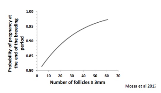
It is believed that AMH concentrations in young animals may be a potential indicator of their fertility in later life. Alward et al, 2019 stated in their review, that AMH is a valuable reproductive management tool, linking productive life and various breeding protocol success to AMH concentrations. Many tests were performed on both sheep and dairy cows. Lahoz et al. 2012 conducted a test on ewes at 10 and 14 months to examine the relationship between AMH concentration in prepubertal ewe lambs and their fertility at first and second matings. The results showed that 23% became pregnant on their first mating and 77% on the second. This was a total of 84.2% overall fertility, concluding that their ability to conceive was correlated with their AMH concentration in the prepubertal phase. AMH was higher in those who conceived after the first mating compared to those who conceived after the second mating or conceived at all. In one study done by (Mossa et al. 2012), it was hypothesized that the total number of follicles ≥ 3mm in diameter is positively associated with reproductive performance in dairy cows. From lecture material available to us, we are aware that there are four main stages of follicular maturation which include the primordial follicle, primary follicle, secondary follicle and tertiary follicle. The primordial follicle develops in the ovaries before birth, the primary follicle after birth, the secondary follicle before puberty and the tertiary follicle is a fully formed antrum, these are also called antral follicles. Three hundred and six lactating dairy cows were assessed between two spring and one autumn breeding season. Ovarian ultrasonography was used to determine the Antral Follicle Count (AFC). The results from this test concluded that as the probability of pregnancy at the end of the breeding season increased, the AFC also increased. Cows with a lower AFC, have poorer fertility. A lower number of follicles in the ovaries of an animal means that there are less viable eggs to be fertilised, thus lowering fertility. This is demonstrated on the graphs below.
|
Figure 1.1 |
Figure 1.1 demonstrates the positive correlation between the number of follicles over 3mm in size and the probability of pregnancy at the end of the breeding period.
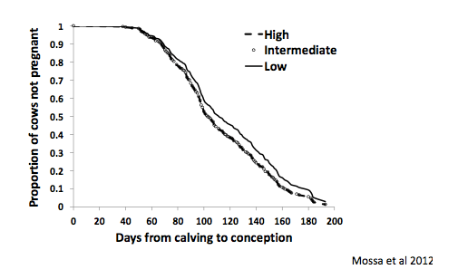
It was noted that dairy cows with a lower AMH level also had a lower conception rate at first Artificial Insemination (AI). A greater number of AI’s were needed to conceive with a longer calving interval in comparison to those with a higher AMH level. This can be seen on the Figure 1.2. During the follicular stage of oestrous, behavioural changes accompany hormonal changes where the dominant hormone is estradiol. The UVMB physiology department advised that some behavioural changes include restlessness and sexual receptivity, suggesting the animal is ready to be artificially inseminated. These behavioural changes vary amongst different species.
|
Figure 1.2 |
In a study done by (Mossa et al. 2017), AFC was hypothesised to be positively associated with the total number of follicles and oocytes in the ovaries of cattle. In order to test this theory, young adult beef heifers were analysed in relation to their AFC level per wave. The ovary opposite the one with the most recent ovulated dominant follicle was removed for analysis. It was weighed and the number of primordial, preantral and antral follicles and oocytes were counted. Ages and bodyweight’s were constant with very little variation. Results showed that those with lower levels of AFC have 60% smaller ovaries and a much lower number of oocytes. A count of fewer morphologically healthy follicles (80%) and oocytes in these animals was also recorded. Animals with low vs high AFC values had 80-90% lower total number of each type of morphologically healthy follicle and oocyte in the ovary.
Maternal Influences on Ovarian Reserve
Nutrition
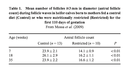
(J.J Ireland et al. 2011) conducted a study which hypothesized that alterations in the maternal diet affect follicular development. He showed this by restricting the intake of beef heifers to 60% of their maintenance energy requirements before conception until the end of the first trimester of pregnancy. The results from this show that, even though the birth weight of calves was unaltered, the AFC concentration of those born to nutritionally restricted dams, was 60% lower than those of the control, as seen in the results below (Figure 2.0).
|
Figure 2.0 |
When (Mossa et al. 2017) tested the same hypothesis, the results were very similar. Female calves born to dietary restricted mothers had an extremely low ovarian reserve. Consistently low AMH levels from the age 4 months until 1.8 years were seen, as well as a low AFC from 7 weeks to 1.6 years of age and increased FSH concentrations. Another experiment conducted by (Sullivan et al. 2010), examined the impact high levels of protein fed to dams had in the second trimester. It resulted in a reduced number of healthy antral follicles in the offspring of these heifers, but unfortunately AMH levels were not measured.
Disease
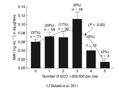
The effects of disease in the dam during pregnancy has also been examined in cattle. It is believed to play a crucial role in the development of the foetus and its health. A recent study conducted by (Mossa et al. 2017) resulted in new data which showed dairy cows with a high SCC measurement exceeding 200,000 may produce female offspring with lowered ovarian functionality and thus reduced fertility in adulthood. The experiment which was performed on 192 Holstein heifers at 12 months of age measured the connection between AMH levels and the size of the corresponding ovaries, total follicle number and oocyte number in the ovary and its function. These results were then correlated with the SCC of the heifer’s dam. The results can be seen in Figure 3.0. The black lines of polynomial regression shows that female offspring of cows with 4 or 5 SCC measurements >200,000 are also recorded to have lower AMH concentrations and smaller ovaries in comparison to those with offspring of cows with 0-3 SCC measurements.
|
Figure 3.0 |
Breed
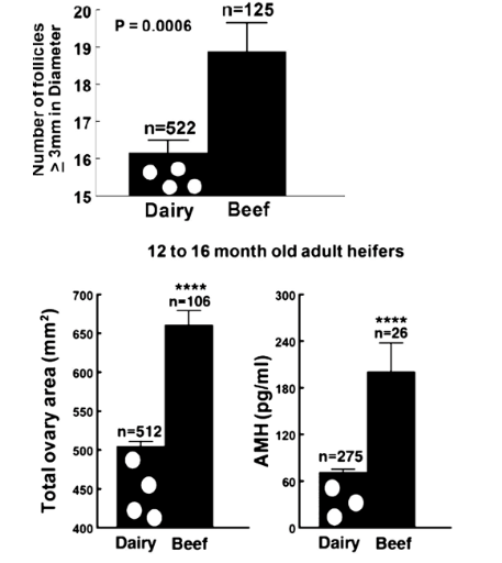
(Mossa et al. 2017) said that beef cattle have higher AMH levels than those of dairy cows. There has not been any reports made on this, although many studies have indicated that the AMH concentration and follicle number may be lower in dairy cows compared to that in beef cattle. In a study done by (Mossa et al. 2017) in the lab, they noted the AMH level, follicle number and ovarian size. This was produced by ultrasonography 96 hours after the prostaglandin treatment. It was conducted on 12-16 month old Holstein dairy heifers and also on Angus x Charolais crossbred beef heifers. A graph can be seen below showing results. These records showed that ovary size, follicle numbers and AMH concentration were lower in the dairy heifers in comparison to the beef heifers. AMH concentration was recorded similarly in prepubertal calves but a significant difference was not found. It is thought that this may be because the blood samples weren’t collected on the same day between beef and dairy calves. Whether or not there is a difference in AMH concentrations between dairy breeds is under debate as an experiment to test this theory between Holstein and Jersey cows conducted by (Pfeiffer et al. 2014) noted no difference. Following this, (Ribeiro et al. 2014) tested a large number of dairy cows and the results concluded that Jersey cows had the highest concentration of AMH. It was also tested on Gyr dairy zebu heifers, Murrah buffalo and Holstein heifers with Gyr having the greatest AMH concentration when compared by (Baldrighi et al. 2014).
In summary of this information found, it can be assumed, but is not yet defined, that AMH concentrations are higher in beef cattle compared to dairy cows and these concentrations differ between genetic dairy groups or breeds.
|
Figure 4.0 |
Figure 4.0 (Mossa et al. 2017), shows the number of follicles ≥3mm in diameter, total ovarian area in mm which was determined using ultrasonography and also AMH concentrations (pg/ml). These tests were recorded approximately 96 hours after prostaglandin F2a treatment in 12-16 month old Holstein dairy and crossbred beef heifers
Conclusion
Fertility
Evidence from all studies show that for AMH measurement, a single blood sample is sufficient to determine relative AFC and quality of follicles and oocytes in the ovaries of animals. From reading (Mossa et al. 2017) paper on variation in the ovarian reserve, strong evidence suggests high variation in the amount of follicles and oocytes in the ovaries, negatively affecting ovary function. These negative effects, may in turn, result in suboptimal fertility in the offspring. The comparison of cattle with a low vs high AFC are as follows; 1) Decreased size of ovary 2) A lower number of morphologically healthy follicles and oocytes 3) A long-standing rise in gonadotrophin secretion with lower AMH and progesterone concentrations during oestrus 4) A reduction in endometrium thickness 5) A higher amount of cell markers for lessened oocyte quality. Many of these phenotypic differences have also been reported in more mature, less fertile cattle in comparison to their younger counterparts. Taking all this evidence into consideration, it is clear that the potential use of AMH concentration is supported as a predictor of fertility in cattle.
Maternal Influence
In relation to disease or illness during pregnancy and its effects on follicle number and the size of the ovarian reserve, there was a large amount of preliminary data collected, presenting us with evidence showing that disease has a negative effect on AMH concentrations (Mossa et al., 2017). As previously discussed, this has a high correlation with the size of the ovarian reserve and thus fertility. However more evidence by experimentation and larger numbers is required to demonstrate this fully. If this were proved, herd fertility as well as overall herd health and milk production could possibly be improved by culling cows which have chronically elevated SCC during pregnancy.
It is evident that certain alterations in the maternal diet affects follicular development, resulting in offspring with decreased AMH levels leading to an extremely low ovarian reserve. For instance, high protein resulted in a reduced level of healthy AFC. Overall, a well-balanced diet is essential for an optimal ovarian reserve.
The analysis of breed specific AMH levels and the ovarian reserve is still unknown. Throughout this essay we analysed the findings of various different scientific papers but discovered that no definite conclusions exist. Therefore, the question whether lower AMH concentrations found in dairy cows in comparison to beef cattle, due to a smaller ovarian reserve, is yet to be explored.
To conclude, it is clear the Anti Mullerian Hormone and the ovarian reserve play a huge role in the fertility of mammals. Although there are many scientific studies surrounding this subject, further research is required to enable AMH to become a predictor of fertility in mature animals. There is detailed research available regarding dairy cows, in comparison to that of cattle or sheep, offering greater potential in the advancement of fertility.
References
- Alward KJ, Bohlen JF. 2019. Overview of Anti-Müllerian hormone (AMH) and association with fertility in female cattle. Reproductive Domestic Animals. 2020 Jan;55(1):3-10.
Baldrighi J, Sá Filho MF, Batista EO, Lopes RN, Visintin JA, Baruselli PS & Assumpção ME. 2014. Anti-Mullerian hormone concentration and antral ovarian follicle population in Murrah heifers compared to Holstein and Gyr kept under the same management. Reproduction in Domestic Animals 49 1015–1020.
Caraviello DZ, Weigel KA, Shook GE & Ruegg PL. 2005. Assessment of the impact of somatic cell count on functional longevity in Holstein and Jersey cattle using survival analysis methodology. Journal of Dairy Science 88 804–811.
Cate RL, Mattaliano RJ, Hession C, Tizard R, Farber NM, Cheung A, Ninfa EG, Frey AZ, Gash DJ & Chow EP. 1986. Isolation of the bovine and human genes for Müllerian inhibiting substance and expression of the human gene in animal cells. Cell 45 685–698.
Ireland JJ, Smith GW, Scheetz D, Jimenez-Krassel F, Folger JK, Ireland JLH, Mossa F, Lonergan P & Evans ACO. 2011. Does size matter in females? An overview of the impact of the high variation in the ovarian reserve on ovarian function and fertility, utility of anti-Mullerian hormone as a diagnostic marker for fertility and causes of variation in the ovarian reserve in cattle. Reproduction, Fertility and Development 23 1–14.
Josso N, Cate RL, Picard JY, Vigier B, di Clemente N, Wilson C, Imbeaud S, Pepinsky RB, Guerrier D & Boussin L. 1993. Anti-müllerian hormone: the Jost factor. Recent Progress in Hormone Research 48 1–59.
- Jost A. 1947. The age factor in the castration of male rabbit fetuses. Proceedings of the Society for Experimental Biology and Medicine 66 302.
Lahoz B, Alabart JL, Monniaux D, Mermillod P & Folch J. 2012. Anti- Müllerian hormone plasma concentration in prepubertal ewe lambs as a predictor of their fertility at a young age. BMC Veterinary Research 8 118.
- Mossa F, Carter F, Walsh SW, Kenny DA, Smith GW, Ireland JL, Hildebrandt TB, Lonergan P, Ireland JJ, Evans AC. 2013. Maternal undernutrition in cows impairs ovarian and cardiovascular systems in their offspring. Biol Reprod. 2013;88(4):92. doi: 10.1095/biolreprod.112.107235.
- Mossa F, Jimenez-Krassel F, Scheetz D, Weber-Nielsen M, Evans ACO, Ireland JJ. 2017. Anti-Müllerian hormone (AMH) and fertility management in agricultural species. Reproduction. 2017;154:R1–11.
Mossa F, Walsh SW, Butler ST, Berry DP, Carter F, Lonergan P, Smith GW, Ireland JJ & Evans AC. 2012. Low numbers of ovarian follicles ≥3mm in diameter are associated with low fertility in dairy cows. Journal of Dairy Science 95 2355–2361.
Pfeiffer KE, Jury LJ & Larson JE. 2014. Determination of anti-Müllerian hormone at estrus during a synchronized and a natural bovine estrous cycle. Domestic Animal Endocrinology 46 58–64.
Ribeiro ES, Bisinotto RS, Lima FS, Greco LF, Morrison A, Kumar A, Thatcher WW & Santos JE. 2014. Plasma anti-Müllerian hormone in adult dairy cows and associations with fertility. Journal of Dairy Science 97 6888–6900.
Sullivan TM, Micke GC, Greer RM & Perry VE. 2010. Dietary manipulation of Bos indicus heifers during gestation affects the prepubertal reproductive development of their bull calves. Animal Reproduction Science 118 131–139.
Figure References:
Ireland JJ, Smith GW, Scheetz D, Jimenez-Krassel F, Folger JK, Ireland JLH, Mossa F, Lonergan P & Evans ACO. 2011. Does size matter in females? An overview of the impact of the high variation in the ovarian reserve on ovarian function and fertility, utility of anti-Mullerian hormone as a diagnostic marker for fertility and causes of variation in the ovarian reserve in cattle. Reproduction, Fertility and Development 23 1–14.
- Mossa F, Jimenez-Krassel F, Scheetz D, Weber-Nielsen M, Evans ACO, Ireland JJ. 2017. Anti-Müllerian hormone (AMH) and fertility management in agricultural species. Reproduction. 2017;154:R1–11.
Mossa F, Walsh SW, Butler ST, Berry DP, Carter F, Lonergan P, Smith GW, Ireland JJ & Evans AC. 2012. Low numbers of ovarian follicles ≥3mm in diameter are associated with low fertility in dairy cows. Journal of Dairy Science 95 2355–2361.

