NEUROTRANSMITTERS AND THEIR CONNECTION TO EMOTIONS, WITH A FOCUS ON DISGUST
Rakonczay, Z. M., Ryan, F. and Sequeira, L. M.
Definition
Disgust has been recognized as a basic emotion since Darwin (1872/1965). Like other basic emotions, disgust has a characteristic facial expression, an appropriate action (distancing of the self from an offensive object), a distinctive physiological manifestation (nausea), and a characteristic feeling state (revulsion) ( Rozin and Fallon, 1987).
Evolutionary Background
Disgust is a deeply ingrained emotion shaped by natural selection, vital to our self-preservation by helping us steer clear of situations that may negatively impact our fitness. There are different types of disgust, notably sexual, moral and pathogenic,which influencing each other and often merge. They are expressed similarly in our initial reaction to things deemed disgusting. Common reflexes include recoiling/retreating, grimacing, turning pale, stomach clenching and occasionally fainting or vomiting. These expressions also help signal to others that something is not right.
At its core, it is a food related emotion and a major contributor to the feeling of pathogenic disgust. The fundamental idea when it comes to food is the “principle of contamination”. For the purpose of this essay contaminants are objects that with even the most minimal contact with previously acceptable food, will make that food unacceptable; the oldest and most universally recognised contaminant being faeces. Food, while being vital for survival, is also one of the main ways for pathogens to enter our bodies, therefore, there are conditions that “food” must meet before being considered acceptable nourishment, and will otherwise be rejected as being non-food, a contaminant or contaminated. It has been noted that nearly all objects considered disgusting are organic, and usually animal related (e.g. vomit, faeces, blood and other bodily fluids) as these pose the most risk to our welfare.
The feeling of contamination not only influences our judgment in food, but also making social decisions. This can be seen in humanity’s treatment of different species.
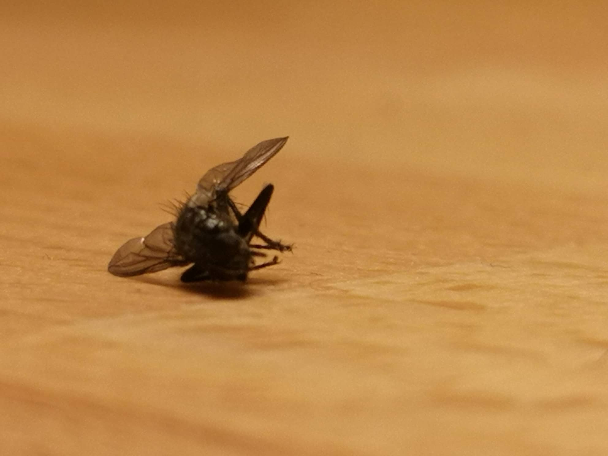
Humans are unwilling to eat different animals for unalike reasons. The most obvious being that they themselves disgust us and are considered contaminants. The easiest way to prove this is by bringing them into contact with food: even a short interaction will make many humans reject the previously acceptable food, or at the very least, reconsider eating it. This leads to us recognising them as pests and therefore care little about their wellbeing, often because they are vectors for pathogens, or suggest that the area is not sanitary (e.g. rats, cockroaches, flies (see Figure 1.)).
|
Figure 1. Example of a common contaminant - the |
Less obvious is that an animal, due to its nature or physical appearance, is appreciated by humans and usually not considered a contaminant. However, this does not mean it is considered acceptable to eat them due to the sense of moral disgust, which becomes engaged as we would feel complicit in the animal’s destruction should we chose to eat it. (e.g. dogs, cats, butterflies, pandas (see Figure 2.)).
|
Figure 2. Non-contaminant |
Moral disgust is vaguer than its pathogenic counterpart, as it is largely based on what individuals deem acceptable or threatening. It influences the kind of people we associate with, how close we want to be to someone, and ultimately, the partners we choose. Numerous factors are at play here: our sense of morality is still influenced by a fear of pathogens, so when judging a potential partner for example, personal hygiene is as much of a factor as their behaviour towards others or their personal views.
Disgust has therefore played a massive role in human history. It has influenced what we eat, therefore how we gather and preserve food so that it stays acceptable, which lead to improving storage and agriculture which made towns and cities possible. As people started living in larger and closer communities, to avoid chaos, moral disgust had to develop, and laws began appearing. At first it was to stop people pathologically disgusting behaviour such as murder, bestiality, and cannibalism. These laws were later refined as civilisations developed further and more complex issues arose. Living closer together also made the need for good hygiene even more crucial, and a lack of it got people and places shunned.
Anatomy and Mechanism
The limbic system of the brain is the primary structure involved in emotional behaviour. The complex sensory information processing occurring in the cortex can directly influence the limbic system. Conversely, limbic processing can strongly influence higher-level cognitive integration occurring in the cortex (Online_Coursebook_'1'). The limbic system can be subdivided into the cortical and subcortical regions. The cortical region is comprised of; cingulate gyri, the piriform lobe and the hippocampus. The subcortical region is comprised of; the habenula of the epithalamus, the hypothalamus and the thalamus of the diencephalon), the interpeduncular and tegmental nuclei of the mesencephalon and the amygdala of the rhinencephalon.
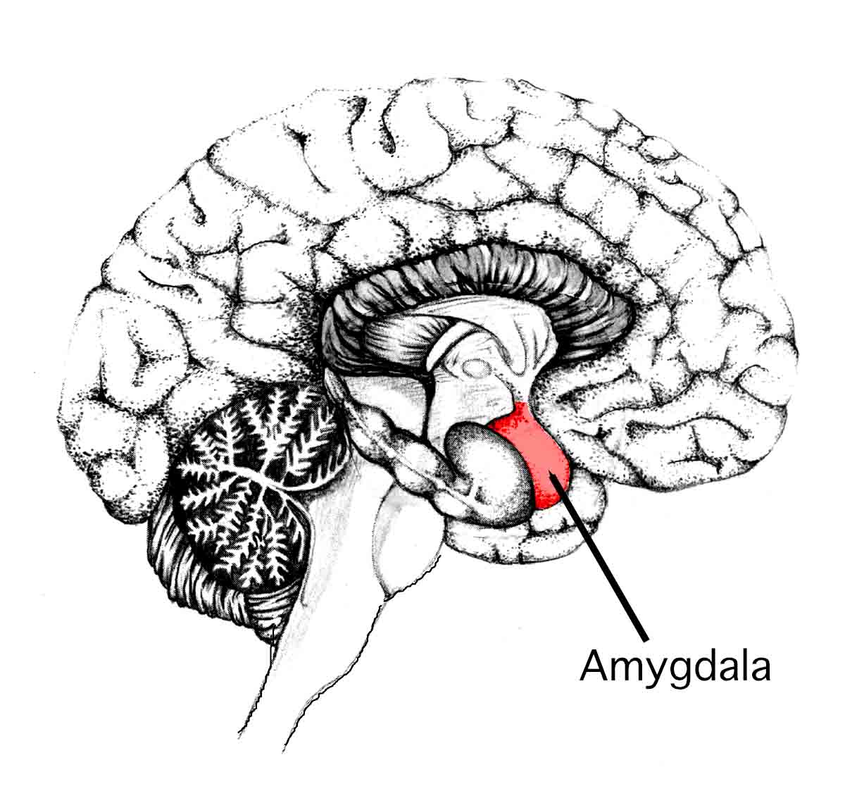
The key anatomical structures associated with emotional behaviour are the amygdala and the hippocampus. As both are highly complex systems, only the mechanism of the amygdala will be discussed in this review (see Figure 3.).
|
Figure 3. The Amygdala |
The amygdala is in the temporal lobe of the brain. Classification is not definitive however classification of the regions can be defined in terms of the nuclei present. The cortical, medial and central nuclei are referred to as the cortico-medial region whilst the lateral, basal and accessory nuclei are referred to as the basolateral region (Online_Article_1'). The amygdala receives various sensory information such as visual, auditory and olfactory from other parts of the brain. It can also receive information from other higher centres such as the prefrontal cortex. The information can either travel via the thalamus to the amygdala or directly to the amygdala (Online_Video_1). The latter results in a reaction without cognitive processing. The stimuli are integrated, analysed and sends an output to initiate a reaction. The function of the reaction is dependent on the part of the brain that receives the output. Connections to and from the amygdala to the other parts of the brain are plentiful. The main connections are by the stria terminalis and the ventral amygdalofugal pathway (Online_Article_1'). Lesions in the amygdala lead to difficulty in associations between stimuli and emotional states. E.g. If prey is aware of predator will result in the feeling of danger which will cause the flight of the prey. Damage to the amygdala will hamper this response. As mentioned previously, there are many neurotransmitters of the amygdala such as serotonin and low levels of dopamine and noradrenaline being the main neurotransmitters associated with disgust. The neurotransmitter is dependent on the connective pathway from the amygdala to the destination in the brain.
The emotion of fear is the most studied emotion researched in reference to the amygdala. The reasoning being that fear is vitally important for survival. It elicits an inhibitory behavioural response. Inhibitory responses are associated primarily with the lateral region of the amygdala. Disgust is another emotion that elicits an inhibitory response therefore parallels can be drawn.
Sensory inputs to the amygdala from the cerebrum mainly terminate in the lateral nucleus. Amygdala outputs tend to originate in the central nucleus, the most peptide-rich region of the brain, and are carried by peptide-containing fibres in the stria terminalis & ventral amygdalofugal pathway. The lateral nucleus is connected to the lateral hypothalamus via the ventral amygdalofugal pathway.
The amygdala has a baseline electrical activity. When a new stimulant is introduced the electrical is rapidly increased. The electrical response decreases with the frequency of exposure to the stimulant. If a stimulant is ambiguous, e.g. binocular rivalry: two different monocular images are displayed to each eye, causing subjective perception to alternate between the two, the electrical activity does not attenuate.
Classical conditioning, or Pavlovian Conditioning, is the pairing of a neutral stimulus with a potent stimulus. If the potent stimulus is no longer present, the negative reaction will decrease and eventually become extinct. This mechanism is known as Extinction. This is an active process. Cognitively the brain recognises the association between the potent and neutral stimulus is no longer valid. The basal nucleus is situated functionally between the lateral nucleus (where most inputs are received) and the central nucleus (when most outputs are generated). The basal nucleus actively inhibits the connection between the lateral and central nuclei thus the negative response is eliminated. The inhibition through the basal nucleus can be removed rapidly thus causing symptoms such as Post Traumatic Stress Disorder (Online_Video_1).
The Neurotransmitters Associated with Disgust
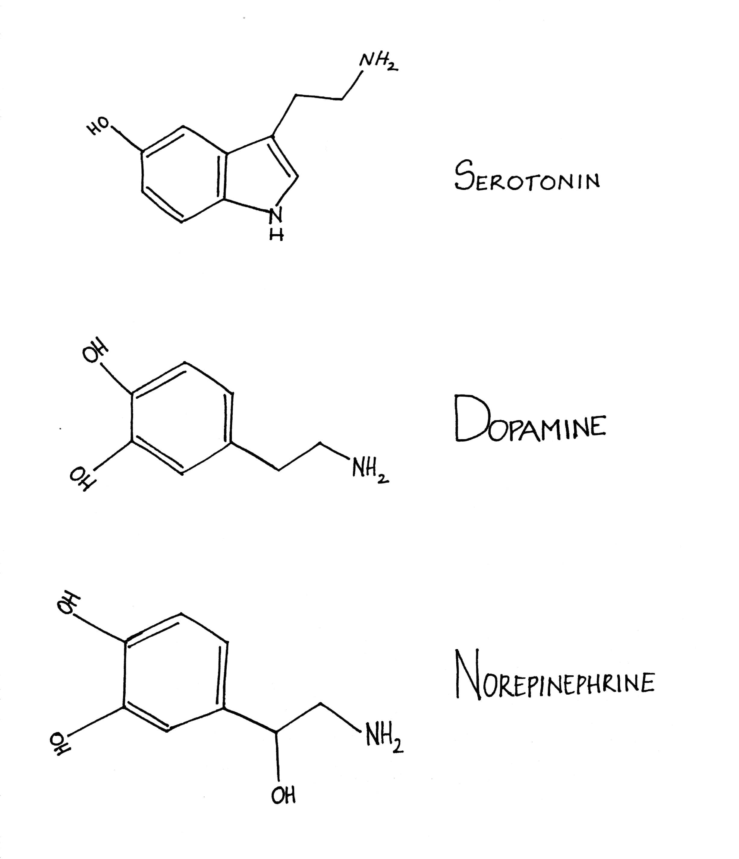
The brain has certain neurotransmitters that release chemicals to provoke an emotion that leads the body of the organism to act a certain way. There are some for disgust as well. High levels of serotonin and low levels of dopamine and noradrenaline are the main neurotransmitters associated with disgust. γ-aminobutyric acid (GABA), glutamic acid and N-methyl-D-aspartate- also neurotransmitters associated with disgust, but in this review the focus is on serotonin, norepinephrine and dopamine (see Figure 4.).
|
Figure 4. The 3 main neurotransmitters of disgust |
Serotonin’s sole precursor is tryptophan and is produced within the gut neurons and enterochromaffin cells on the periphery and within the neurons of the raphe in the brain stem centrally. Most of this tryptophan is bound to plasma albumin, so it is unavailable for transport to the brain, but can be taken up by the brain upon release. In the central nervous system (CNS), L-tryptophan is hydroxylated to 5-hydroxytryptophan by the enzyme tryptophan hydroxylase type-2, which is followed by a decarboxylation with the enzyme l-aromatic acid decarboxylase to serotonin (5-hydroxytryptamine, 5-HT).
There are multiple serotonin receptors from 5-HT1-5-HT7. Large areas of the human brain contain serotonergic neurons that innervate it. Most of these projections arise from neuronal cell bodies in the dorsal and median raphe and neighbouring nuclei of the lower brain stem. Projections reach to the hippocampus, amygdala, hypothalamus, thalamus, neocortex, and basal ganglia, although most structures receive some serotonergic innervation. This is a very thorough and vital network since serotonin modulates crucial functions such as sleep, control of appetite and temperature. An imbalance in levels of serotonin can lead to several psychiatric disorders, the most common of which are depression and anxiety. To combat this, a group of agents known as SSRI (Selective Serotonin Reuptake Inhibitors) are used. A well know SSRI is Escitalopram, which has a quick onset response with a few side effects. An enantiomer of it, Citalopram can also be used intravenously in animals.
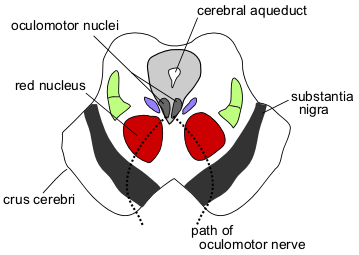
Dopamine and Norepinephrine/Noradrenaline are less abundantly produced and their low levels are associated with disgust. Dopamine neurons are found in the midbrain’s substantia nigra and ventral tegmental area (see Figure 5.). They project heavily to the dorsal and ventral striatum, respectively. In contrast, noradrenergic axons, originating from the brain stem locus coeruleus (nucleus in the pons) and medulla project diffusely to most brain regions, including those innervated by the dopamine and norepinephrine system. Overlapping occurs in areas between the dopamine and norepinephrine systems like the hypothalamus, bed nucleus of the stria terminalis (BNST), and frontal cortex. Stress and emotion-related information is processed here.
|
Figure 5. The Midbrain |
Although disgust is an emotion that comes naturally and consciously for human beings, for research purposes, it may also be conditioned. According to Tuerke et al. (2012), “The forebrain structure critical for the production of nausea-induced conditioned disgust may be the insular cortex (IC), which is an area involved in the sensation of nausea in humans and other animals” (Kaada, 1951; Penfield and Faulk, 1955; Fiol et al, 1988; Contreras et al, 2007; Catenoix et al, 2008).
In the case of rats, according to Tuerke et al. (2012), in his findings from the work of Breslin et al. (1992) and Parker (1995) it is said that disgust reactions can be defined by the gaping (large openings of the mouth and jaw, with lower incisors exposed), which is characterized as the most sensitive and predominant conditioned disgust reaction. Certain experiments were conducted on Male Sprague Dawley rats in which they were prevented from producing conditioned gaping reactions (as a response to disgust) by infusing them with a saline paired saccharin solution (Parker, 1998), in turn to try to deplete the effect of 5-HT, i.e.- serotonin. It is known through experiments conducted mentioned in the work of Vicario (2014) that “cocaine blocks the reuptake of serotonin and other neurotransmitters into presynaptic neurons by binding to the neuronal membrane transporters for this monoamine.(Ritz et al.,1990) and “suppresses serotonin synthesis which leads to decreased levels of 5-hydroxyindoleacetic acid (5-HIAA) (Baumann et al.,1993).
Serotonin is important in executing disgust’s conditioned functions. It has been shown that the depletion of forebrain serotonin (5-HT) by 5,7-dihydroxytryptamine (5,7-DHT) lesions leads to the prevention of induced conditioned disgust reactions such as “gaping,” to “taste related” aversive stimulation (Limebeer et al.,2004).
Disgust can also be prominent in mental illnesses where the patient can be averse to normal conditions such as in the psychiatric disorder, Anorexia Nervosa (AN) in which the patient has a marked disgust for food and starve the body of nutrients, therefore putting their health at risk due to an imbalance in their neurotransmitters.
Nature vs. Nurture
Like most emotions, disgust is subjective, and developed by an individual’s conditioning, surroundings, and biological instincts, leading to a wide variation of what is considered disgusting amongst different sexes, age groups and cultures.
Since disgust as an emotion is mostly developed through conditioning, it is not present in most young children so they cannot yet differentiate between safe and dangerous ingestible items. This is particularly dangerous during infancy when children explore the world mainly through putting objects in their mouths, so they depend on their guardians to ensure they avoid interacting with dangerous materials. This guidance is enhanced and corrected through social interactions with their agemates who often label certain actions as disgusting. People feel ashamed when labeled as disgusting, and learn quickly to avoid the habits that gave them that label.
The difference between the sexes go back to the basic human family structure. Women evolutionarily have been tasked with raising children and ensuring their safety, so have to be more aware of pathogens and other dangers like antisocial or threatening behaviour, therefore a lower disgust threshold is prevalent. Men on the other hand, had the role of a hunter and fighter.
A higher tolerance was needed as a contaminant is blood, and therefore a higher tolerance is advantageous.
What are the results (observed) of disgust?
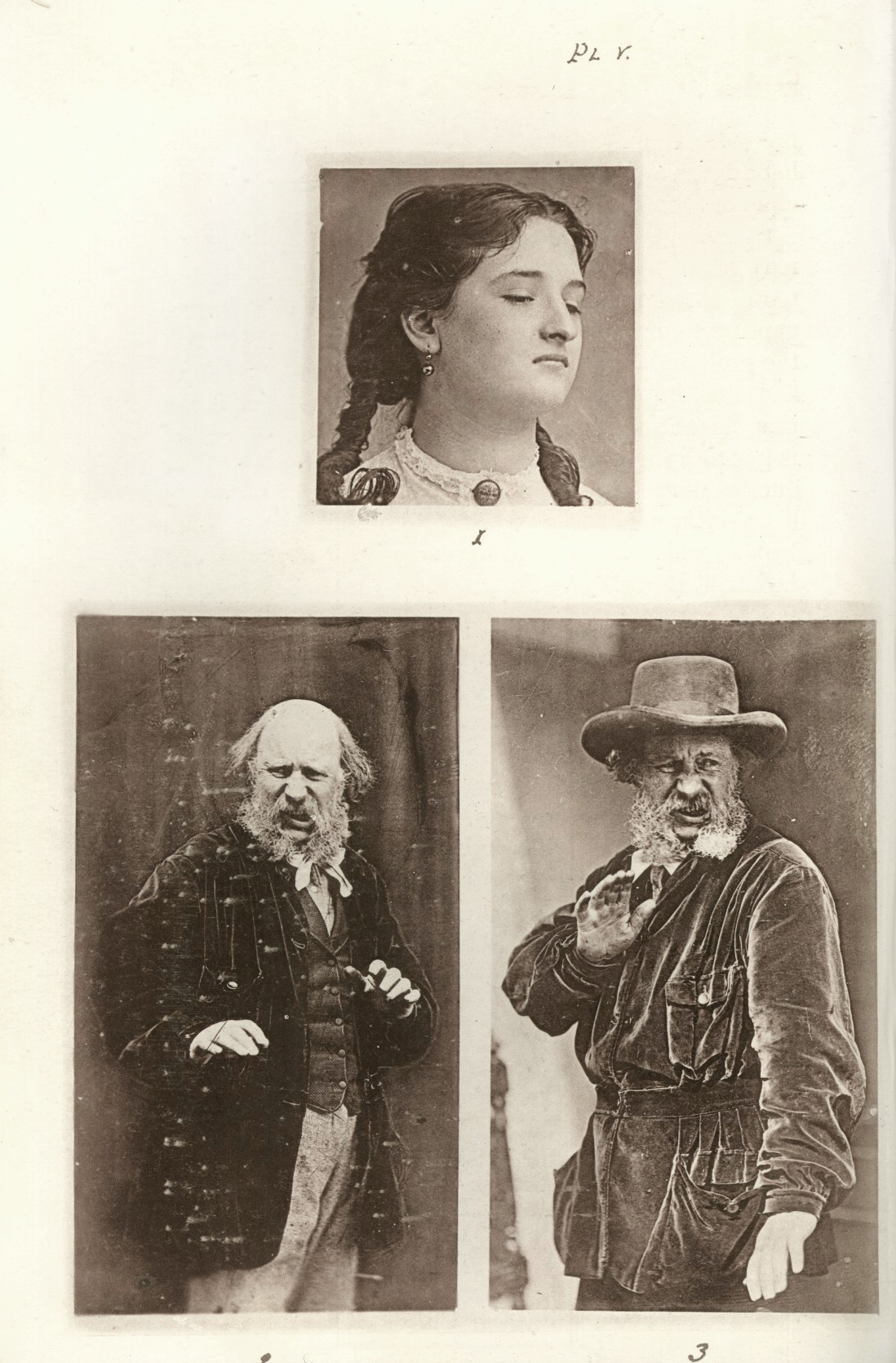
Usually, when exposed to a substance that elicits disgust, one’s facial expression would resort to frowning, said substance would be avoided, certain sounds could be made to show the feeling of unwantedness, body language would resort to that of wanting to move away and staying far from this particular substance. Common body language seen in disgust is baring of the teeth, lower of the eyebrows, tightening the eyelid, and wrinkling the nose (see Figure 6).
|
Figure 6. Depiction of Darwinian Emotions |
However, facial expressions can also be miscategorized if proper context is not accompanied with it. This is known as the confusability effect (Hassin et al.,2013)
It is often seen that disgust and anger can be can be confused if the disgust facial expression was shown in an angry context because of their similarities (Susskind et al.,2007). Whereas with fear, disgust in a fear context was perceived correctly since the two are not that similar in action (Aviezer et al.,2008). A good example of that would be seeing a rat in the house. It is seen as an unwanted rodent and possibly diseased, so people are usually disgusted by it, as well as feared for personal reasons.
Conclusion
While it does provoke very negative feelings, disgust is a vital emotion enabling us to live in a functional and safe society, and helping us avoid danger. Being a socially influenced emotion, it is a very subjective based on a person’s background and age, and may adapt to the environment a person finds themselves in. It is influenced by serotonin, dopamine and noradrenaline, 3 powerful neurotransmitters, which make it a very strong emotion, difficult to ignore and potentially if an imbalance may form, it may lead to mental illnesses such as but not limited to anorexia, anxiety and depression. Being such an old and ingrained emotion, it is easy to forget that it is one which has guided the judgement of our environment, our animals and each other, and has lead humankind to live in the societies we know today.
References
References from Journals
Alves-Neto, W.; Guapo, V.; Graeff, F.; Deakin, J.; Del-Ben, C. (2010): Effect of escitalopram on the processing of emotional faces. Brazilian Journal of Medical and Biological Research, 43 (3), 285-289. doi:10.1590/s0100-879x2010005000007
Aviezer, H.; Hassin, R. R.; Ryan, J.; Grady, C.; Susskind, J.; Anderson, A.; Moscovitch, M.; Bentin, S. (2008): Angry, Disgusted, or Afraid? Psychological Science, 19 (7), 724-732. doi:10.1111/j.1467-9280.2008.02148.x
Baumann,M. H.; Raley, T. J.; Partilla, J. S.; Rothman, R. B. (1993): Biosynthesis of dopamine and serotonin in the rat brain after repeated cocaine injections: a microdissection mapping study. Synapse, 14, 40–50 doi:10.1002/syn.890140107
Breslin, P. A.; Spector A.C.; Grill, H.J. (1992): A Quantitative Comparison of Taste Reactivity Behaviors to Sucrose before and after Lithium Chloride Pairings: A Unidimensional Account of Palatability. Behavioral Neuroscience, 106 (5) 820–836., doi:10.1037/0735-7044.106.5.820
Catenoix, H.; Isnard, J.; Guénot, M.; Petit, J.; Remy, C.; Mauguière, F. (2008): The role of the anterior insular cortex in ictal vomiting: A stereotactic electroencephalography study. Epilepsy & Behavior, 13 (3), 560-563. doi:10.1016/j.yebeh.2008.06.019
Contreras, M.; Ceric, F.; Torrealba, F. (2007): Inactivation of the Interoceptive Insula Disrupts Drug Craving and Malaise Induced by Lithium. Science, 318 (5850), 655-658. doi:10.1126/science.1145590
Curtis, V. (2011): Why Disgust Matters. Philosophical transactions of the Royal Society of London. Series B, Biological sciences: 366 (1583), 3478–3490. doi:10.1098/rstb.2011.0165
Fiol, M. E.; Leppik, I. E.; Mireles, R.; & Maxwell, R. (1988): Ictus emeticus and the insular cortex. Epilepsy Research, 2 (2), 127-131. doi:10.1016/0920-1211(88)90030-7
Hassin, R. R.; Aviezer, H.; Bentin, S. (2013): Inherently ambiguous: Facial expressions of emotions, in context. Emotion Review, 5, 60 – 65.http://dx.doi.org/10.1177/1754073912451331
Kaada, B. R. (1951): Somato-motor, autonomic and electrocorticographic responses to electrical stimulation of rhinencephalic and other structures in primates, cat, and dog; a study of responses from the limbic, subcallosal, orbito-insular, piriform and temporal cortex, hippocampus-fornix and amygdala. Acta Physiol Scand Suppl 24: 1–262.
Limebeer, C. L.; Parker, L. A.; Fletcher, P. J. (2004): 5,7-Dihydroxytryptamine Lesions of the Dorsal and Median Raphe Nuclei Interfere With Lithium-Induced Conditioned Gaping, but Not Conditioned Taste Avoidance, in Rats. Behavioral Neuroscience, 118 (6), 1391-1399. doi:10.1037/0735-7044.118.6.1391
Parker, L. A. (1995): Rewarding drugs produce taste avoidance, but not taste aversion. Neuroscience & Biobehavioral Reviews, 19 (1), 143-151. doi:10.1016/0149-7634(94)00028-y
Penfield, W.; Faulk, M. E. (1955): The Insula. Brain,78 (4), 445-470. doi:10.1093/brain/78.4.44
Ritz, M.; Cone, E.; Kuhar, M. (1990): Cocaine inhibition of ligand binding at dopamine, norepinephrine and serotonin transporters: A structure-activity study. Life Sciences, 46 (9), 635-645. doi:10.1016/0024-3205(90)90132-b
Rozin,P. ; Fallon, A. E. (1987): A Perspective on Disgust. Psychological Review: 94. 23-41 doi:10.1037//0033-295X.94.1.23
Rozin, P.; Ruby, B. M. (2019): Bugs Are Blech, Butterflies Are Beautiful, but Both Are Bad to Bite: Admired Animals Are Disgusting to Eat but Are Themselves Neither Disgusting nor Contaminating. Emotion. 10.1037/emo0000587.
Susskind, J. M.; Littlewort, G.; Bartlett, M. S.; Movellan, J.; Anderson, A. K. (2007): Human and computer recognition of facial expressions of emotion. Neuropsychologia, 45, 152–162. http://dx.doi.org/10.1016/j.neuropsychologia.2006.05.001
Tuerke, K. J.; Limebeer, C. L.; Fletcher, P. J.; Parker, L. A. (2012): Double Dissociation bBtween Regulation of Conditioned Disgust and Taste Avoidance by Serotonin Availability at the 5-HT3 Receptor in the Posterior and Anterior Insular Cortex. Journal of Neuroscience, 32 (40), 13709-13717. doi:10.1523/jneurosci.2042-12.2012
Tybur, J. M.; Bryan, A. D.; Lieberman, D. L.; Caldwell Hooper, A. E.; Merriman, L. A. (2011): Sex Differences and Sex Similarities in Disgust Sensitivity. Personality and Individual Differences, 51, 343-348.
Vicario, C. M. (2014): Aberrant Disgust Response and Immune Reactivity in Cocaine-Dependent Men Might Uncover Deranged Serotoninergic Activity. Frontiers in Molecular Neuroscience, 7. doi:10.3389/fnmol.2014.00007
Miscellaneous References
Konig, H. E., Liebich, H-G. (2014): Veterinary Anatomy of Domestic Animals. Stuttgart, Germany: Schattauter GmbH
Online_Article_1, LeDoux, J. E. (2008): Amygdala: http://www.scholarpedia.org/article/Amygdala
Online_Article_2, CBC (2017) :The Nature of Things: Science, Wildlife and Technology. (n.d.). Retrieved from https://www.cbc.ca/natureofthings/features/the-seven-universal-emotions-we-wear-on-our-face
Online_Coursebook_1, THE NEUROBIOLOGY OF EMOTION. Neural systems, the amygdala, and fear: http://www.neuroanatomy.wisc.edu/coursebook/neuro5%282%29.pdf
Online_Video_1, Jordan, I. (2016): The amygdala: From structure to function: https://www.youtube.com/watch?v=FXMsV-iIzj4
Figure Index
Figure 1 - Picture taken by 3703/E (Rakonczay, Z. M., 2019)
Figure 2 - Picture taken by 3703/E (Rakonczay, Z. M., 2019)
Figure 4 - Drawn by 3628/E (Sequeira, L. M., 2019)

