Epithelial Function and Repair
The most common type of cell within the body is the epithelia,these cells are arranged in sheets and cover most organs within the body, including the skin, respiratory tract and digestive tract. Epithelia in these systems offer the first line of defense to the outside world, we will discuss digestive and respiratory epithelia, which are important for defense. The location within the body, and the function that the epithelia will perform has an impact upon them. These cells can be one or more cell layers and will be highly differentiated depending upon the function performed. These vital functions performed have many specialised mechanisms that will maintain the correct functioning and physiology of the cells. Upon an injury to the epithelia, whether from disease or accidental injury, there is physiological damage. Thus, repair will involve the healing of damaged tissue to restore normal functioning. The damaged tissue will be repaired by the resealing of the epithelial surface barrier to preserve normal homeostasis. The continuity of the epithelia is reestablished in three key mechanisms:
- Epithelia cells from the neighboring area will migrate to the injury site to cover the denuded area, epithelial restitution will occur, this will not require cell proliferation.
- The second step requires cell proliferation;
- Thirdly maturation and differentiation of the undifferentiated epithelium will take place.
This ensures the maintenance of various activities within the epithelium. The main aim is for the body to repair the damaged tissues either with scar tissue, or via regeneration (Strum et Dignass, 2008).
The Digestive System
An Overview of the Gut and its Epithelia
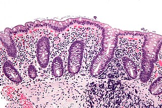
The gut is divided into the oesophagus, stomach, intestine and colon. The oesophagus is composed of stratified squamous epithelia, very similar to that of the epidermis. The stomach, intestine and the colon are composed of simple epithelia, which is one cell layer. This contains different cell lineages in various parts of the gut (Keymeulan et Blanpain, 2012). The corpus of the stomach houses parietal cells, which secretes acid for digestive processes. The mucosa here will secrete a protective barrier that will renew rapidly (Keymeulan et Blanpain, 2012). The small intestines consists of many proliferating crypts, the crypts give rise to villi. These are large protrusions of the epithelium present within the gut lumen. Villi will enlargen the surface area for absorption. Intestinal tissues are one of the quickest proliferating tissues within the body, it will die by anoikis and will completely self-renew within a week (Keymeulan et Blanpain, 2012).

Figure 1: Shows a high magnification micrograph of intestinal spirochetoso, intestinal spirocytes (colonic spirocytes, colonic spirochetes, rectal spirochetosis and rectal sirochetes). H&E stain, a hyperchromatc fuzz on the luminal aspect of the epithelial cells – at brush border can be seen at high magnification.
Major Functions of the Digestive Epithelium
The digestive epithelium is a major barrier between the ‘outside’ and ‘inside’ world. The main functions of the epithelium are to absorb nutrients, ions and water, which is key to sustain life. However, this can be troublesome as the epithelium must balance the above processes whilst protecting the ‘inside’ world from many harmful pathogens, toxins irritants and bacteria that will exists within the lumen of the gut (Raybould, 2012). The health of an individual, whether it be animal or human, will depend upon efficient digestion and absorption of nutrients from food via the gut, this must be done by balancing delivery of food, digestion and absorptive capacity of intestines. The components of a meal entering the epithelium are sensed by gut enteroendocrine cells, thus activating pathways, which will include neural and humoral pathways regulating the gastrointestinal motor. Secretory and absorptive functions also play a role in maintaining plasma glucose levels. The mucosa barrier will be well maintained, as break down of the epithelial barrier can cause many problems varying from gut inflammation and bowel diseases (Raybould, 2012).
Specialised Gut Epithelial Cells
Some gut epithelial cells are specialised enteroendocrine cells that act as sensors, they can detect ions, nutrients, bacterial products and other luminal contents. The epithelial cells are pluripotent, being able to synthesize, store and release many polypeptide hormones. These hormones are essential and can affect different functions within the gut itself via paracrine actions. They can also act as hormones at different sites, including the exocrine and endocrine pancreas and the central nervous system (Raybould, 2012). Enteroendocrine cells will express TLRs, as do many epithelial cells. Many publications have shown this and it was demonstrated in immunohistochemical studies that TLRs in human and murine intestinal tissue that TLR-1, TLR-2 and TLR-4 were expressed on subpopulations of epithelial cells with enteroendocrine cell morphology located within crypt in the colon and small intestines. The receptors will have ligands that cause secretion of cholecystokinin (CCK) from the cells. The mechanism for this is dependent upon MyD88 and protein kinase C, which are intracellular signaling molecules (Raybould, 2012).
Gut Epithelial Repair
Following an injury the gut epithelium will be restored via a rapid migration of the intestinal epithelial cells across the denuded area, this process in known as wound healing and will restore the original integrity of the epithelia. The process has a sequence of events that includes; restitution, proliferation and differentiation of the intestinal epithelia, (see figure 2) (Parlato & Yeretssian, 2014).

Figure 2: Shows the sequence of events involved in epithelium repair that includes; restitution, proliferation and differentiation.
Restitution will be stimulated by several regulatory peptides, growth factors and cytokines. There is also a key input from nucleotide-binding and oligomerization domain-like receptors in the regulation of intestinal inflammation. It begins within minutes, epithelial cells lining the surface will de-differentiate and begin to remodel the cytoskeleton. Actin will reorganise and provide a structural framework that defines the shape and polarity of the epithelia cells, thus pushing the cell forward. The process is regulated by RHO family of guanine triphosphatases (Parlato & Yeretssian, 2014). Proliferation will increase the pool of enterocytes and aims to re-establish the barrier integrity and function. There are many factors that promote tissue repair, one possible is the signal transduction pathways, EGF and TGF- α, that induce restitution and proliferation. It has recently been shown in studies that the Wnt-5a-expressing stromal cells are key for the regeneration of intestinal tissues (Parlato & Yeretssian, 2014).
The epithelial Toll-like receptors (TLRs) will be the main defense against pathogens, engaging the TLR signaling will preserve intestinal epithelial barrier and protect the intercellular gap junctions and promote the expression of various proliferative and anti-apoptotic factors (Parlato & Yeretssian, 2014).
The Respiratory System
Lung Epithelial Function
The mammalian airways can be divided into two parts on the basis of the two main functions; the conducting and the respiratory airways. The conducting airway is composed of the nose, trachea, and bronchi, and is responsible for the transport of air to the lungs. The air is warmed before entering the lungs, and particles are filtered out by cilia. The respiratory airway consists of respiratory bronchi and alveoli, which are lined by epithelium, allowing gas exchange to occur. The epithelium in the lungs is pseudostratified, containing several different cell types, with different morphology and functions (Hollenhorst et al, 2011)

Figure 3: Scanning electron microscope image of the trachea epithelium. There are both ciliated and non-ciliated cells in the epithelium.
The respiratory tract has a vast cell surface area which is in direct contact with inhaled air, exposing the host to different gases and exogenous factors. The epithelium is very complex, it mediates gas exchange, clearance of mucus by cilia, defense of the host, and homeostasis of the surfactant in order to maintain lung sterility and stability. In adults, the lungs are not mitotically active. However, the epithelium can proliferate rapidly after an injury, in order to maintain proper structure and function of the lung (Park et al, 2005).
A healthy lung is able to remain sterile without recruiting phagocytes, due to mucociliary action and antimicrobial polypeptides in the surfactant that covers the epithelium. Inhaled bacteria are trapped in the mucus covering the airways, and are propelled upwards towards the larynx by ciliary movement. This process is completed by antibacterial polypeptides, these are secreted from the epithelium and sub-mucosa glands. Some pathogens are resistant to the polypeptides, but they still do not cause inflammation, which emphasizing the importance and the efficiency of the mucociliary action. If these mechanisms fail, lung diseases such as cystic fibrosis can develop (Moskwa et al, 2006). Studies have shown that extra-pulmonary cells migrate into the lung from the bone marrow, and play a role in the repair of respiratory epithelium after injury. Progenitor cells are also thought to participate in epithelial repair, however, not as rapidly. Another cell type which is considered important in cell repair is the non-ciliated Clara cells which maintain a high proliferative capacity, due to the large quantity of cells participating in epithelial repair, resulting in a rapid and widespread growth of the lung (Park et al, 2005).
Lung Epithelial Injury
Injury to the lung epithelium can be a result of bacterial or viral infections, inflammation, allergic reactions (asthma), exposure to xenobiotics (e.g., cigarette smoke), pathology of unknown origin (idiopathic fibrosis), physical trauma (mechanical ventilation), or cancer (Crosby & Waters, 2010). Depending on the nature of the injury, lung repair mechanisms begin immediately and initiate an acute inflammatory response involving cytokine release, immune cell recruitment, and activation of the coagulation cascade. Repair of respiratory epithelium after injury is critical in order to recover homeostasis (Crosby & Waters, 2010). Injury results in the release of factors that help repair mechanisms including:
- epidermal growth factor and fibroblast growth factor families (TGF, KGF, HGF)
- chemokines (MCP-1)
- interleukins (IL-1, IL-2, IL-4, IL-13)
- prostaglandins (PGE2)
These factors unite processes to promote cell spreading and migration.
During the repair process, progenitor cells are recruited from circulation, and together with local progenitor cells, they multiply and undergo phenotypic differentiation in order to restore the stability and functionality of the epithelial layer (Crosby & Waters, 2010). Within the lungs non-ciliated Clara cells are found, they remain unchanged for the most part of their life, however, an injury will trigger proliferation. Pulse-chase experiments have been used to show that these cells are able to proliferate, and that they were free of secretory granules and smooth endoplasmic reticulum. When the injured tissue is replenished, the Clara cells are inactivated until they are needed again (Quilhac & Sire, 1999).

Figure 4: Transmission electron microscope of a thin section through the bronchiolar epithelium of the lung (mouse), which consists of ciliated cells and non-ciliated cells (Clara cells). The image shows ciliary microtubules in transverse and oblique section. In the apex re basal bodies that anchor the ciliary axonemes.
Clara cells provide the lung with a remarkable repair capacity, even if regeneration is slow. Basal and Clara cells are found in the conducting airways and type II cells within the alveoli, these cells aid maintenance of the lungs proliferative capacity. Type II cells can regenerate lung tissue in the case of pneumonectomy. If an injury occurs via hyperoxia or sulphur dioxide, the Clara and type II cells will be activated and initiate proliferation (Park et al, 2005).
Maintaining Lung Homeostasis
To ensure correct functioning of the lung, there is a rapid cell response during the repair of the epithelium. During the repair permeability barriers have to be maintained and restored, proliferation initiated, and finally differentiation of the cells found within the epithelium of the lung. The genome will be influenced by signals produced via growth factors, cytokines and transcription factors, thus initiating morphogenesis, differentiation, and pulmonary homeostasis (Park et al, 2005). Transcription factors will regulate the expression of specific genes, which initiate the differentiation of cellular groups found within the adult lung. These factors will also regulate homeostasis of the surfactant, transport of fluids, clearance of mucus by cilia, defense, and other processes crucial for pulmonary homeostasis (Park et al, 2005).
Pathways Involved in Epithelial Repair in the Airways
Injury stimulates activation of macrophages and the recruitment of immune cells. Mesenchymal stem cells or niche stem cells are involved. Soluble factors are secreted by immune and non-immune cells acting on epithelial cells and fibroblasts. Spreading and migration, closing the wound and reestablishing intact barrier function. TGF-β signaling results in trans-differentiation of epithelial cells to myo-fibroblasts and the initiation of a chronic inflammatory response. During resolution, immune cells disappear from the area of injury, macrophages are deactivated, cells differentiate, and hyper-proliferation is reduced through apoptosis This allows tissue remodelling (Crosby & Waters, 2010)
Importance of Epithelia
Approximately 1% of gut epithelial cells are specialised enterendocrine cells, which are capable of detecting ions, nutrients, bacterial products and many luminal contents. These cells are pluripotent, and are capable of synthesizing and releasing polypeptide hormones Gut hormones play a critical role within uptake of nutrients within the stomach and small intestines (Raybould, 2012). There has been exciting progress in understanding signal transduction mechanisms, which govern mucosal wound healing after an injury (physical, chemical or bacterial). Intestinal tissue repair is a finely tuned process which involves, regulatory peptides, growth factors, cytokines and innate immune recognition (Parlato & Yeretssian, 2014). Many principles regarding lung cell differentiation and proliferation are derived from developing studies. The mechanisms controlling the formation and repair of this cellular and functional diversity are relatively unknown at present. Current studies provide evidence that ciliated cells participate in repair of the bronchiolar epithelium after acute injury, through rapid squamous metaplasia and re-differentiate into mature, columnar cell types. Ciliated cells are capable of remarkable plasticity, undergoing dynamic changes in cell shape and gene expression during repair (Park et al, 2005).
References
Crosby L. M., Waters C. M. (2010): Epithelial repair mechanisms in the lung. American Journal of Physiology - Lung Cellular and Molecular Physiology 298:(6) L715–L731. Complete Article
Eisenhauer P., Earle B., Loi R., Sueblinvong V., Goodwin M., Allen G. B., Weiss D. J. (2013): Endogenous Distal Airway Progenitor Cells, Lung Mechanics, and Disproportionate Lobar Growth following Long-Term Post-Pneumonectomy in Mice. Stem Cells 31:(7) 1330–1339. Complete Article
Hollenhorst M.I., Ritcher K., Fronius M. (2011): Ion Transport by Pulmonary Epithelia. Journal of Biomedicine and Biotechnology 2011: Article ID 174306. Complete Article
Keymeulan A., Blanpain C. (2012): Tracing epithelial stem cells during development, homeostasis, and repair. The Journal of Cell Biology 197:(5) 575-584. Complete Article
Miki T., Lehmann T., Cai H., Stolz D.B., Strom S.C. (2005): Stem cell characteristics of amniotic epithelial cells. Stem cells(Dayton, Ohio) 23:(10) 1549-59. Complete Article
Moskwa P., Lorentzen D., Excoffon K.J., Zabner J., McCray P.B. Jr, Nauseef W.M., Dupuy C., Bánfi B. (2006): A Novel Host Defense System of Airways Is Defective in Cystic Fibrosis. American Journal of Respiratory and Critical Care Medicine 175:(2) 174-83. Complete Article
Park K., Wells J., Zorn A., Wert S., Laubach V., Fernandez L., Whitsett J. (2006): Transdifferentiation of Ciliated Cells during Repair of the Respiratory Epithelium. American Journal of Respiratory Cell and Molecular Biology 34:(2) 151-157. Complete Article
Parlato M., Yeretssian G. (2014): NOD – Like Receptors in Intestinal Homeostasis and Epithelial tissue Repair. International Journal of Molecular Sciences 15: 9594-9627. Complete Article
Quilhac A., Sire J.Y. (1999): Spreading, proliferation, and differentiation of the epidermis after wounding a cichlid fish, Hemichromis bimaculatus. The Anatomical Record 254:(3) 435-51. Complete Article
Raybould H. (2012): Gut microbiota, epithelial function and derangements in obesity. The journal of physiology 590:(3) 441-446. Complete Article
Strum A., Dignass A. (2008): Epithelial restitution and wound healing in inflammatory bowel disease. World Journal of Gastroenterology 14:(3) 348-353. Complete Article
Other References
Figure 1: http://commons.wikimedia.org/wiki/file:intestinal_spirochetosis_-_high_mag.jpg
Figure 2: http://www.vivo.colostate.edu/hbooks/pathphys/digestion/stomach/gibarrier.html
Figure 3: http://commons.wikimedia.org/wiki/file:lung_epithelium_80294-2.6.jpg
