Endocrinology of the horse, hormonal changes during and after physical activity
Contents
-
Endocrinology of the horse, hormonal changes during and after physical activity
- Introduction:
- Pituitary Gland
- Adrenal Gland
- Kidneys:
- Heart:
- Investigation 1: Adrenocorticotropin, cortisol and catecholamines response to various exercises.
- Investigation 2: Equine Plasma β-endorphin concentrations are affected by exercise intensity.
- Pancreatic endocrinology:
- Investigation 3: Neuroendocrine control of blood volume, blood pressure and cardiovascular function in horses
- Investigation 4: Insulin Sensitivity of Horses during different exercise intensities.
- Conclusion:
- BIBLIOGRAPHY
Introduction:
The compound word Endocrine originates from “endo”, meaning internally and the Greek term “krinein” which means to distinguish or separate. Endocrine system controls the internal environment to optimize cell functions through hormone release. A hormone is a substance secreted and produced by organs or glands and is carried through circulation to a specific organ causing local physiological response. Especially in horses where they spend most of their time exercising for long period of time, body responses due to hormone secretions occur.(Mckeever, 2002).
Pituitary Gland
The hypophysis is an endocrine gland located at the basal part of the brain and consists of three different lobes from which numerous hormones are secreted. The lobes include the posterior lobe also known as the neurohypophysis, the anterior lobe or the adenohypophysis and the intermediated lobe. All three named according to their anatomical position in the brain. The effect and intensity of secretion of the hypophyseal hormones where investigated during resting and exercising states (Mckeever, 2008)
Adenohypophysis (Anterior lobe)
Adrenocorticotropin hormone (ACTH):
The ACTH release is regulated by the corticotropin releasing hormone (CRH) which is produced by the hypothalamus and is inhibited by glucocorticoids. The secretion of ACTH is stress dependent and stimulates cortisol secretion. A large precursor protein named proopiomelanocortin is synthesized and facilitates in ACTH synthesis. The function of ACTH is to regulate cortisol levels which is a steroid and glucocorticoid hormone released from the adrenal gland (Bowen, 2018; Mckeever, 2008; Nagata et al, 1999).
Endorphins:
Endorphins are opiate-like peptides that regulate pain sensation(suppressors) or pleasure. They are released from the adenohypophysis of the pituitary gland during stressful conditions, such as exercising. Sometimes they are classified as neurotransmitters. They are quite useful substances in allowing the horse to execute high intensity exercises (Mckeever, 2008; Mehl et al, 1999).
Neurohypophysis (Posterior Lobe)
Arginine Vasopressin (AVP):
A neurohypophysis hormone that is linked with chronic and acute defense of blood pressure, plasma volume and also for fluid and electrolyte homeostasis. The secretion of AVP carried out by osmoreceptors in the hypothalamus and baroreceptors in the atria of the heart. Some of AVP functions include Vasoconstriction and decrease of free water clearance (McKeever and Hinchcliff, 1995).
Adrenal Gland
The adrenal glands are paired and located near the kidneys. They consist of two layers one of them being the adrenal cortex and the other the adrenal medulla. The medullary region produces adrenaline and noradrenaline, and the cortical region produces a variety of steroid hormones that are divided to three categories:
1. Mineralocorticoids like aldosterone
2. Glucocorticoids such as cortisol
3. Gonadotocorticoids for instance androgens and estrogens
Aldosterone:
Aldosterone has a crucial role in electrolyte homeostasis, mainly for sodium and potassium. Its target organ is mostly the kidneys, elevating the sodium and chloride reabsorption as well as potassium excretion. Aldosterone effects can be exerted in the digestive tract as well (intestines), regulating the uptake of electrolytes and water. Stimulation of aldosterone secretion involves the drop of sodium and increases plasma hydrogen and potassium ions. Effects of aldosterone changes with different exercise intensities can affect the horse’s long-term reabsorption of sodium and water by the kidneys and the intestinal tract (McKeever and Hinchcliff, 1995).
Cortisol:
The lowest levels of cortisol in the blood plasma occur at late evening and night, on the other hand the levels of cortisol in the mornings are the highest. Cortisol is characterized as an ester hormone and produced by the effects of stressors. Cortisol has two main functions. Substrate mobilization and immune modulation. During exercise cortisol enhances gluconeogenesis and lipolysis. Glucose utilization is decreased and protein catabolism increases thus increasing the release of amino acids used during exercise as a source of energy. Tissue repairment occurs by the amino acids released (Mckeever, 2008). Cortisol has an anti-inflammatory effect and suppresses immune reactions. The rates of cortisol release are affected by the duration and intensity of the exercise. For this reason, the levels of cortisol in the blood can be used as an indicator for the amount and duration of exercise the horse was subjected to (Mckeever, 2008).
Catecholamines:
The release of catecholamines is altered by the alarm reaction or the “Fight or Flight” response. The main catecholamines are epinephrine (adrenaline) and norepinephrine (noradrenaline). They are produced by the adrenal medulla. Catecholamines exert their effects through specialized receptors that are divided to two categories. Alpha and Beta adrenergic receptors which are further divided to α -1, α -2, β -1 and β -2. The secretion of catecholamines increases in response to increasing exercise intensity. When the release of catecholamines is extreme the heart rate and cardiac output increases. By causing vasoconstriction, blood flow to tissues that will not aid the animal in the alarm reaction, decreases. In this way blood is directed away the digestive system towards the muscles (Mckeever, 2008).
Kidneys:
Renin (Plasma renin activity – PRA):
Hormone secreted by the juxtaglomerular apparatus and is responsible for the synthesis of angiotensinogens to angiotensin-I which is further converted to angiotensin-II in the lungs. Its main function is vasoconstriction and also the secretion stimulation of the mineralocorticoid aldosterone. Different change of the renin angiotensin system is linked to the maintenance of blood pressure during vigorous exercises (McKeever and Hinchcliff, 1995).
Heart:
Atrial natriuretic peptide (ANP):
Hormone produced by the heart that is crucial in the maintenance of blood flow and blood pressure in the duration of the exercise. ANP is stored as granules in the atrial walls and secreted during atrial contractions. This hormones functions as a vasodilator and important for natriuresis. ANP suppresses the effects of Vasopressin, renin and aldosterone secretion and aldosterone binding to the tubules of the kidney, thus acting as an antagonist to these hormones. ANP exerts its effects by receptor mechanisms found in a variety of organs such as neurohypophysis, adrenal cortex, heart and lungs (McKeever & Hinchcliff, 1995)
Investigation 1: Adrenocorticotropin, cortisol and catecholamines response to various exercises.
In a study carried by Tsuruta-Cho, Utsunomiya, and Tochigi in the equine research institute in Tokyo, Japan the concentration of cortisol, catecholamines (adrenaline, noradrenaline) and ACTH in the blood in response to various exercises was studied. In this experiment three healthy stallions and two mares of five years old with body weight around 450kg were used in three exercise styles. A catheter was inserted to the right jugular vein of each animal to investigate hormonal concentrations changes in the blood during and after the exercise (Nagata et al, 1999).
The exercise protocol consisted of two forms of exercise using a treadmill:
An incremental exercise involving a 5-minute warm up by the horses at a speed of 4m/s at 0% incline. Then the treadmill incline was set to 10% and the initial speed was 2m/s. The velocity of the treadmill was increasing every minute by 1m/s. The exercise was terminated when the horse reached complete exhaustion. The recovery period involved the horse walking at 1m/s for 15 minutes at 0% incline (Nagata et al, 1999).
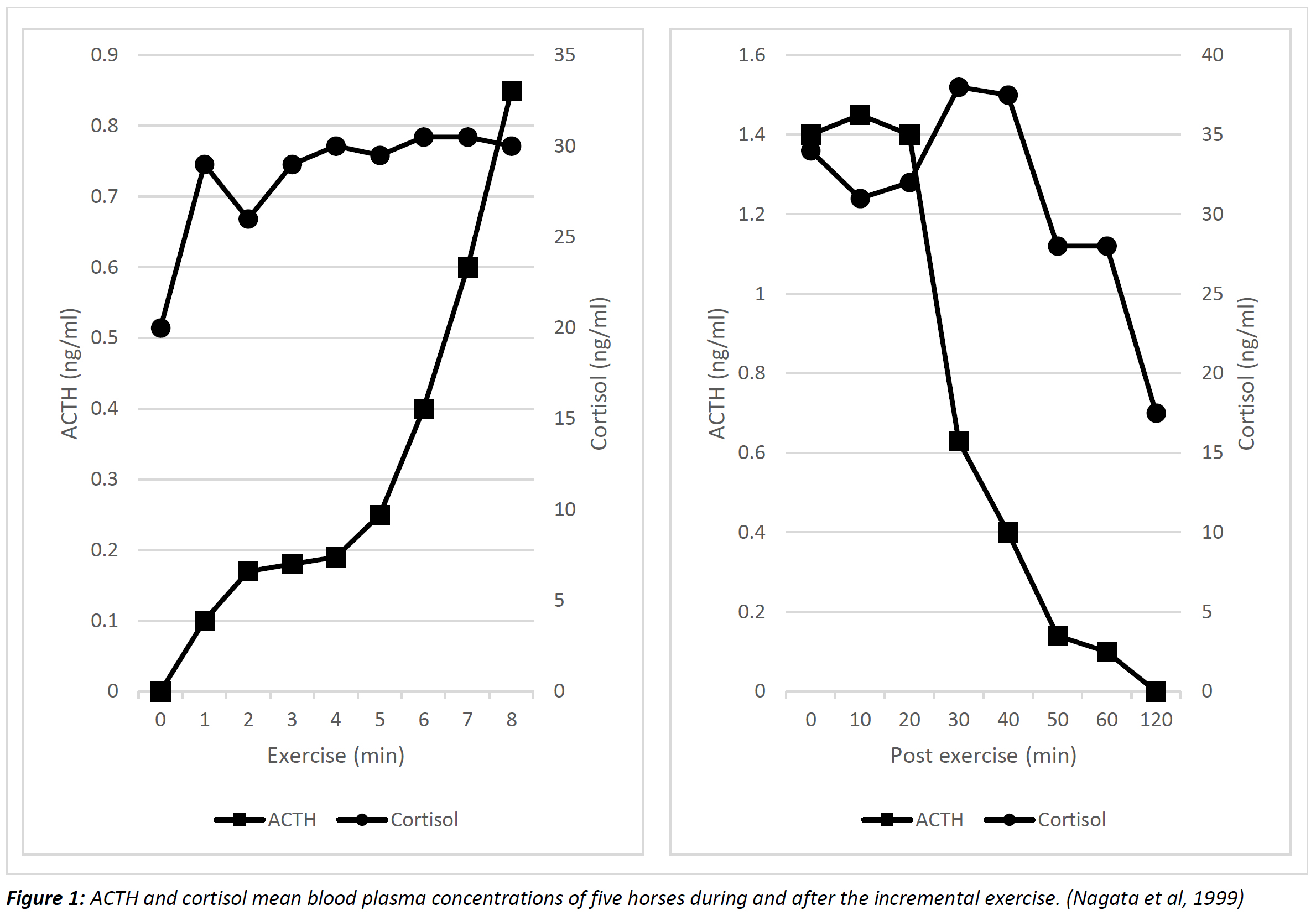
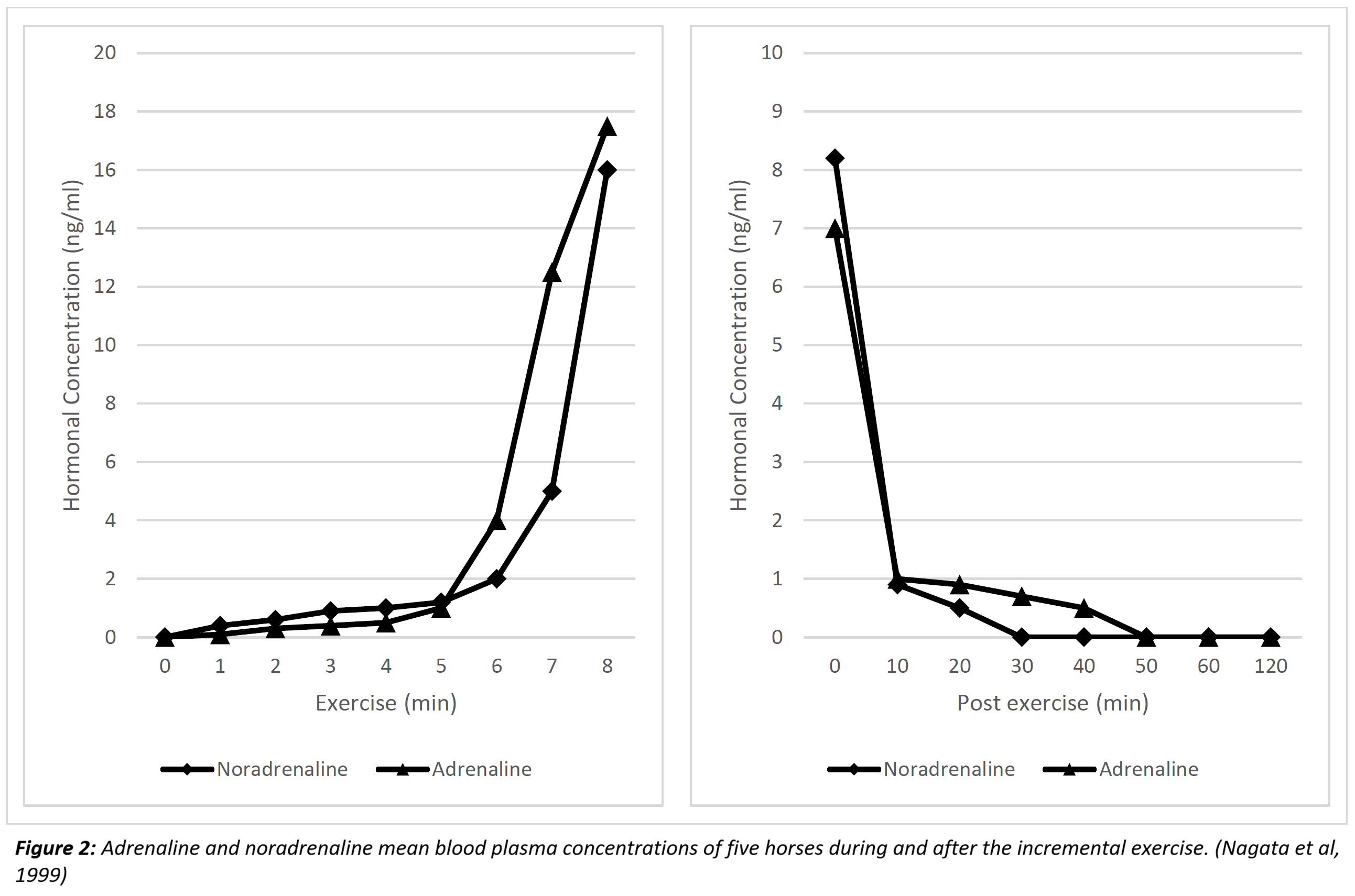
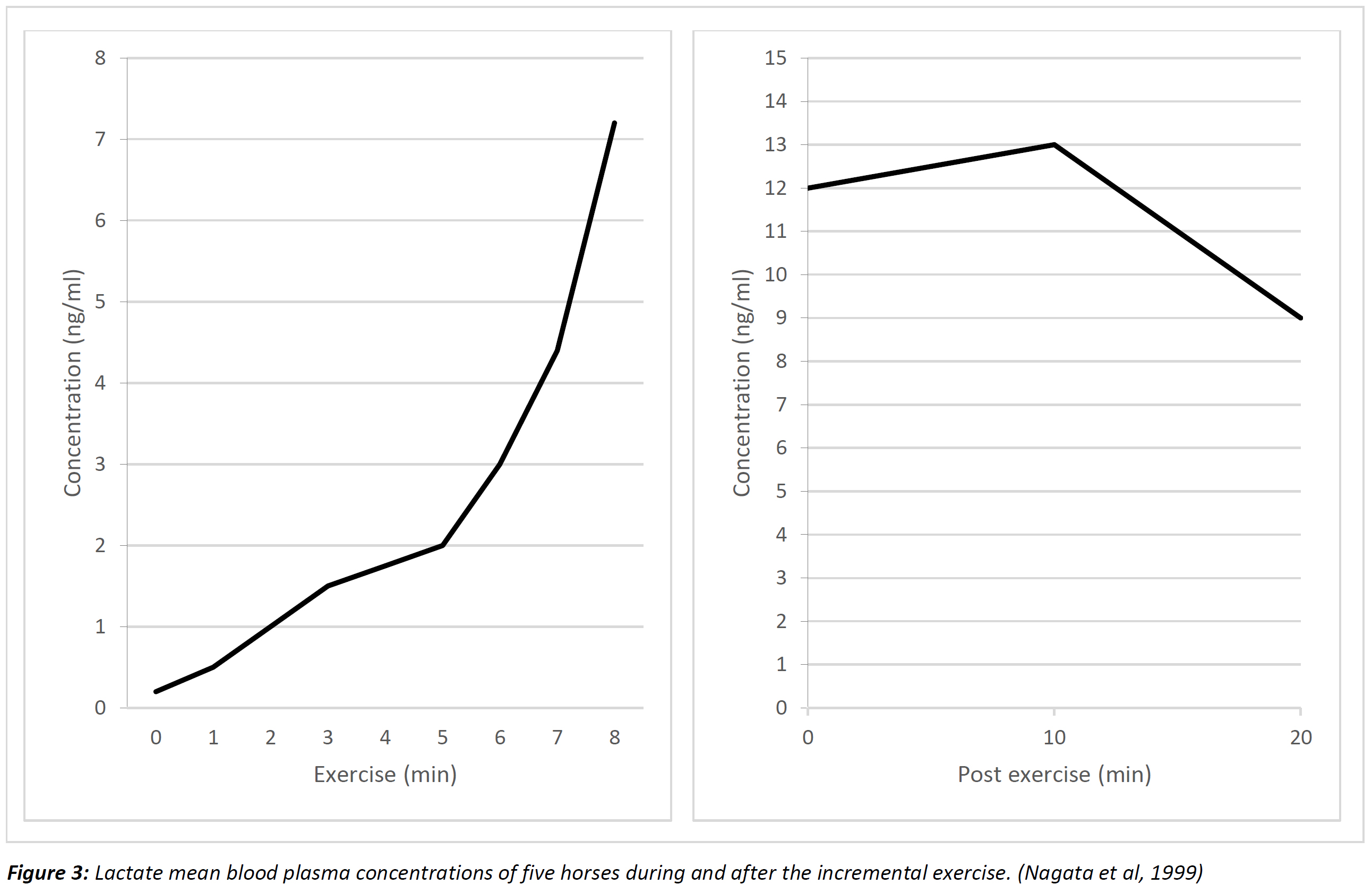
Figure 1 to 2 show the hormonal concentration changes in the horses during and after the incremental exercise. The concentration of the ACTH increased drastically during the first minute of exercise as shown in Figure 1. As the exercise duration was increasing, the concentration of adrenaline, noradrenaline and ACTH was increasing as well. As shown in Figure 1 the ACTH mean concentration in the blood was increased from 0 to around 33 ng/ml. However, the concentration of the cortisol in the blood during the increase of the above-mentioned hormones was relatively constant after the initial increase of around 0.25 ng/ml (Nagata et al, 1999).
Noradrenaline and adrenaline concentrations during the first five minutes of exercise showed a slight elevation (Figure 2). After the first 5 minutes of exercise the concentrations of the hormones were increased dramatically during a short period of time (Nagata et al, 1999).Levels of lactate formation were also measured as an indicator to identify the point at which the horses came to an exhaustion (Nagata et al, 1999). These are indicated in Figure 3.
A relative workload exercise where the treadmill velocity was adjusted in order for the horses to reach 105% and 80% maximum oxygen consumption whereas the incline was kept constant at 10% (Nagata et al, 1999).
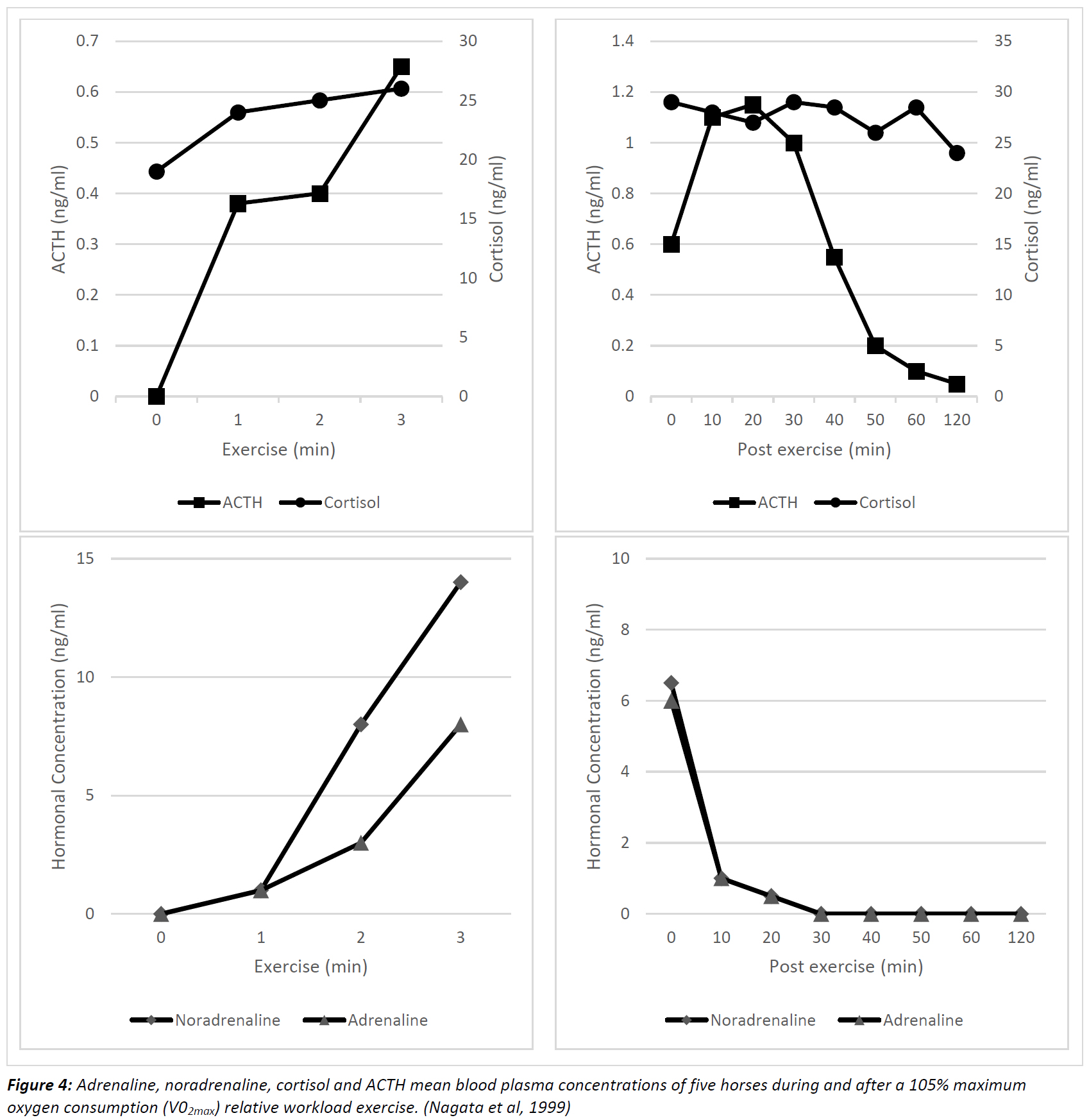
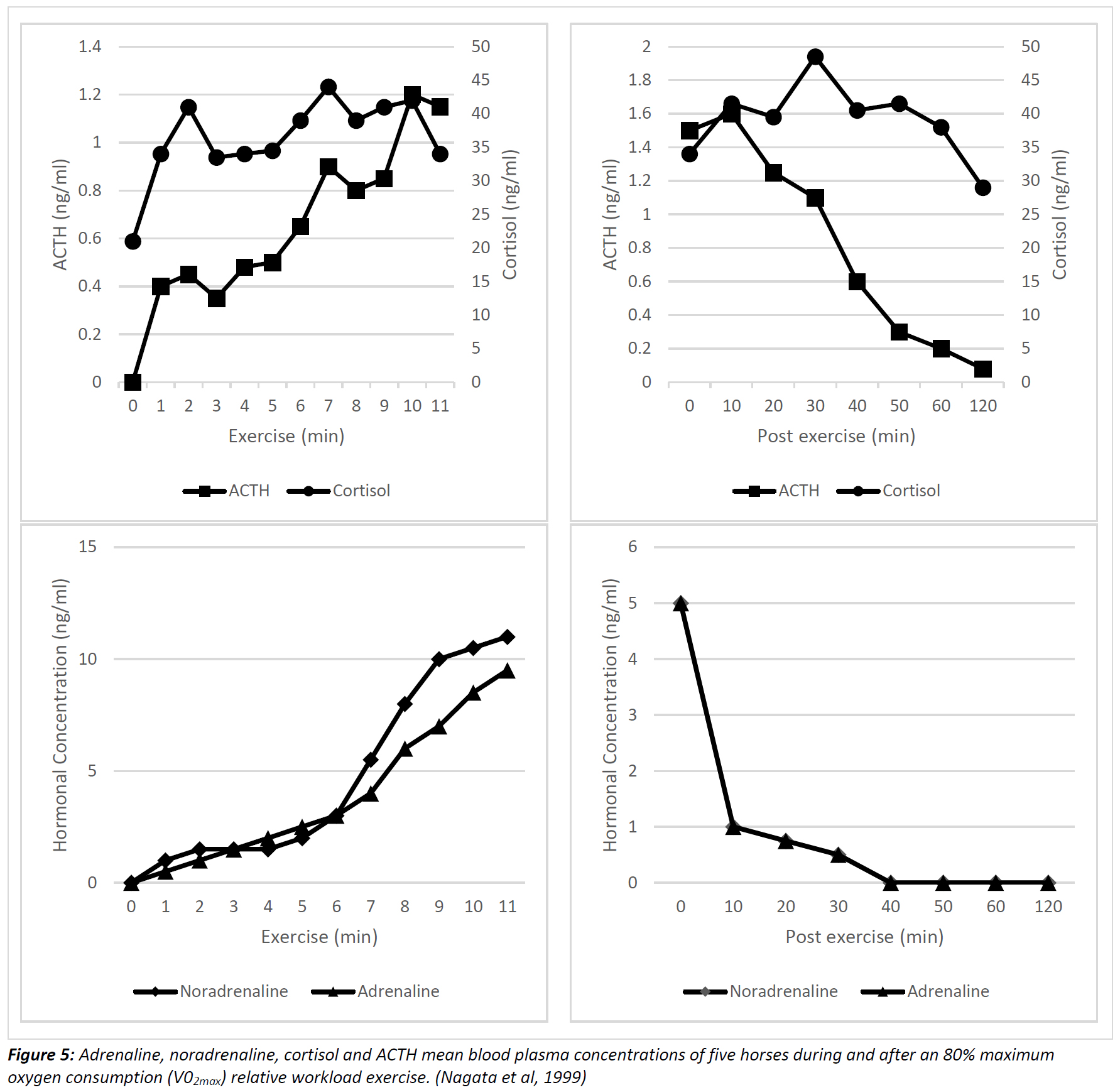
Figures 4 and 5 show that at 105% maximum oxygen consumption (V02max) all 4 hormones showed a linear increase in concentrations. The maximum values of cortisol and ACTH were reached 15 minutes after the exercise was terminated. However, for adrenaline and noradrenaline the maximum concentration values were reached just after the completion of the exercise (Nagata et al, 1999). At 80% maximum oxygen consumption the ACTH concentration was increasing together with velocity on the other hand cortisol concentration was maintained relatively constant after the initial increase (Nagata et al, 1999).A similar behavior in adrenaline and noradrenaline concentrations to the incremental type of exercise was observed at 80% maximum oxygen consumption, in the relative workload exercise protocol (Nagata et al, 1999).
Conclusion:
To sum up the study proves that secretion of the hypophyseal and adrenal hormones are influenced by different intensities of exercise. Also, a positive correlation is shown between lactate and hormonal concentrations. It is clearly indicated by the results of the study that the blood plasma cortisol concentration is associated with the duration of the exercise. In addition, the study can provide information about the stress levels acquired by the horses during the different forms of exercise studied (Nagata et al, 1999).
Investigation 2: Equine Plasma β-endorphin concentrations are affected by exercise intensity.
This experiment was carried out by Mehl, M. L. et al (1999) with a sample of eight mares (5 thoroughbreds and 3 crossbreds) of ages 3 to 13 years old and of average body weight of 450kg.
A warm up exercise was carried prior by the horses. The warm up exercise consisted of 5-minute walking and 5-minute trotting at a speed of 4m/s (Mehl et al, 1999). The horse’s maximal oxygen consumption was determined a week prior to the experiment.
The horses were exercising for 20 minutes in order to reach 60% or were allowed to exercise until exhaustion with a maximum oxygen consumption monitored steady at 95% (Mehl et al, 1999).
Investigators applied a catheter in the left jugular vein of the horses to examine plasma concentrations of beta endorphins (Mehl et al, 1999).
Results:
It is proven by the results, that there is a smaller increase of beta endorphin concentration in the blood plasma prior to the exercise until the end of the warm up. There is a much steeper elevation in the concentration of beta endorphins after the warm up until the end of the second part, the vigorous exercise (20 minutes duration) (Mehl et al, 1999). The results are summarized in table 1.

Pancreatic endocrinology:
The pancreas is an exocrine V-Shaped organ that follows alongside the duodenum. Digestive enzymes and bicarbonate are secreted by the acini cells of the pancreas to the duodenum via the pancreatic duct. This can be described as the digestive functionality of the pancreas. The pancreas also contains islets of Langerhans which are classified into 4 types of cells:
1. α-cells which produce glucagon
2. β-cells which produce insulin
3. δ-cells which produce somatostatin
4. γ-cells which produce pancreatic polypeptide
The most important hormones related to exercise and physical activity are insulin and glucagon which function in glucose metabolism (Mckeever, 2008; Thomas, 2000).
Insulin:
Insulin functions as a glucose storage hormone since it transports glucose to be taken up by the cells, hence stimulating glycogenesis. When the horse is exercising glucose is taken up by the muscles independently without the use of insulin. Insulin is very important when the horse recovers after intense exercise when glucagon activity is most active (Mckeever, 2008; Thomas, 2000).
Glucagon:
Glucagon is an insulin antagonist and a catabolic hormone. It stimulates gluconeogenesis and inhibits glycogenesis. It increases glucose and fatty acid concentration in blood plasma. It is used for substrate mobilization and its secretion increases during muscle activity. Glucagon helps the horse maintain endurance by preventing muscle fatigue (Mckeever, 2008; Reece & Campbell, 2002).
Investigation 3: Neuroendocrine control of blood volume, blood pressure and cardiovascular function in horses
According to McKeever and Hinchcliff (1995) the increase of Atrial Natriuretic Peptide (ANP), Arginine Vasopressin (AVP), Plasma Renin Activity (PRA) and aldosterone (ALDO) by exercising are important factors for cardiovascular homeostasis. This is ensured by mechanisms such as blood volume, arterial pressure and plasma tonicity. Researches showed that plasma ANP rises with exercise in horses. It rises in a linear way by strengthening the work intensity from 9±1 pgml at rest to 63±14pg/ml at 9m/s. Throughout stable situation submaximal exercise, plasma ANP rise from 11±1pg/ml at rest to 40±9pg/ml (264%) at 40 min and remained at that level along 60 min of effort (McKeever and Hinchcliff, 1995)
Hormones studied:
Arginine Vasopressin hormone (AVP)
Extracellular tonicity stimulates AVP release. AVP is released during activity above threshold level concentrations. AVP also constricts the vessels, therefore it regulates the blood pressure during exercise. In exercising horses, AVP rises (1min at 4, 5, 6, 7, 8, 9 and 10 m/s) from 4.0 ± 3.0 pg/ml at rest to 95 ± 5.0 pg/ml at speed of 10 m/s as reported in an experiment. Another research showed that during steady state submaximal exercise the AVP rises, because AVP release is inhibited during excess volume. AVP liberation is also correlated to intensity and duration exercise. Intensity correlation is curvilinear explained by an exponential function (r=0.92) (McKeever and Hinchcliff, 1995).
Plasma Renin Activity (PRA)
Researches obtained PRA in horses at rest, after activity, during balance state exercise or after short maximal exercise and proved the relation of PRA increase with intensity exercise (McKeever and Hinchcliff, 1995).
Aldosterone (ALDO)
The drop-in plasma Sodium ion concentration or rise in plasma Hydrogen ion concentration, plasma ACTH and/or increased PRA, causes the release of aldosterone. The study of McKeever and Hinchcliff (1995) identifies factors that prompts the aldosterone release while exercising of horses. A research study proves that the drop of plasma Sodium concentration doesn't affect aldosterone release. However, PRA causes the aldosterone release while it generates Angiotensin I and II; stimulators of aldosterone release (McKeever and Hinchcliff, 1995). Horses running on a treadmill were tested to identify the correlation between ALDO concentration and exercise intensity. There was an elevation of ALDO concentration simultaneously with PRA and specifically PRA had less increase than ALDO which shows that more factors affect ALDO release. Another factor increasing ALDO release is the increase of plasma Potassium concentration. During light activity of the horse a study reported that Sodium release was low for one hour. These are homeostatic requirements for electrolyte homeostasis, therefore aldosterone plays a minimal role during exercise, even though it remains high for hours after exercise (McKeever and Hinchcliff, 1995).
Investigation 4: Insulin Sensitivity of Horses during different exercise intensities.
This case study tests how insulin sensitivity changes as a result of different exercise intensities for a long period of time. Insulin sensitivity is described as how sensitive the body cells are in response to insulin. If the body cells are very sensitive to insulin it means that they can use glucose in the blood more effectively, thus reducing the blood glucose levels eliminating additional insulin release by the pancreas. Insulin sensitivity is referred as insulin resistance (Turner et al, 2011).
In the case study which lasted one year, 31 stable horses went through 3 exercise protocols or a mixture of the three types throughout these months. The three types of exercise were:
1. Turnout (Horses were free in the field and allowed to walk and explore the area without human intervention – Least exercise intensity).
2. Light exercise.
3. Moderate exercise.
The purpose of the study was to determine the intensity of the exercise training required to affect insulin sensitivity. The horses which had a common diet of high insoluble but low soluble carbohydrates. Blood was sampled every month and the insulin concentration was recorded (Turner et al, 2011).
Table 2 summarizes the monthly physical activity carried out by the horses for the duration of the case study.

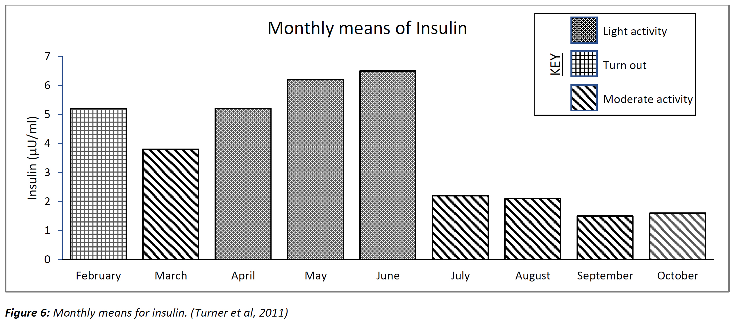
Figure 6 shows the average values of insulin concertation in the blood of the horses
Results:
Insulin concentration was higher in months where horses received only turnout, than in months where horses underwent light exercise. It was the lowest with moderate exercise intensity. Insulin sensitivity was lower in turnout horses than lightly exercised horses and also lower than moderately exercised horses. Insulin sensitivity was higher in moderately exercised horses in comparison with lightly exercised horses. Moderate exercise intensity performed five days per week seems to benefit insulin sensitivity, but an exercise with both light and moderate intensity is recommended to improve insulin sensitivity (Turner et al, 2011).
Conclusion:
Taking everything into consideration, all hormones described above place an important role in the endocrine system during exercise. Carbohydrates and free fatty acids contribute as energy stores to avoid frequent fatigue. Glucagon has a crucial role in homeostasis of blood glucose levels during exercise. In addition, each hormone is influenced differently during exercise. Catecholamines, increase the rate of glycogenolysis, thus enhancing stamina and prevents excessive energy loss. Glucagon and Catecholamines release are influenced by the blood concentration of cortisol. Cortisol triggers gluconeogenesis and protein breakdown to amino acids. These amino acids are useful in repairing muscle, during rest, after exercise, and replace enzymes in the metabolism.
BIBLIOGRAPHY
Bowen, R. (2018): Adrenocorticotropic Hormone (ACTH, Corticotropin).
Mckeever, K. H. (2008): Endocrine Alterations in the Equine Athlete. In Equine Exercise Physiology - The Science of Exercise in the Athletic Horse: (1) 283–284.
McKeever, K. H.; Hinchcliff, K. W. (1995): Neuroendocrine control of blood volume, blood pressure and cardiovascular function in horses. Equine Veterinary Journal 27: (18 S) 77–81. https://doi.org/10.1111/j.2042-3306.1995.tb04894.x
McKeever, K. H. (2002): The endocrine system and the challenge of exercise. Veterinary Clinics of North America - Equine Practice 18: (2) 321–353. https://doi.org/10.1016/S0749-0739(02)00005-6
Mehl, M. L.; Sarkar, D. K.; Schott II, H. C.; Brown, J. A.; Sampson, S.; Bayly, W. M. (1999): Equine plasma β-endorphin concentrations are affected by exercise intensity and time of day. Equine Veterinary Journal 30: (S30) 567–569. https://doi.org/10.1111/j.2042-3306.1999.tb05285.x
Nagata, S.; Takeda, F.; Kurosawa, M.; Mima, K.; Hiraga, A.; Kai, M.; Taya, K. (1999): Plasma adrenocorticotropin, cortisol and catecholamines response to various exercises. Equine Veterinary Journal, 30: (S30) 570–574. https://doi.org/10.1111/j.2042-3306.1999.tb05286.x
Reece, J.; Campbell, N. (2002): Biology. San Francisco: Benjamin Cummings.
Thomas, M. N. (2000): Essentials of Human Physiology (First). Gold Standard Multimedia.
Turner, S. P.; Hess, T. M.; Treiber, K.; Mello, E. B.; Souza, B. G.; Almeida, F. Q. (2011): Comparison of Insulin Sensitivity of Horses Adapted to Different Exercise Intensities. Journal of Equine Veterinary Science 31: (11) 645–649. https://doi.org/10.1016/j.jevs.2011.05.006
Table 1: Mean ± s.e. plasma beta-endorphin concentrations (pg/ml) in 8 horses before (pre-exercise), following a warm up, and after 20 min exercise at 60% VO2max or at fatigue following exercise at 95% VO2max. (Mehl et al, 1999).
Table 2: Monthly physical activity. (Turner et al, 2011)
Figure 1: ACTH and cortisol mean blood plasma concentrations of five horses during and after the incremental exercise. (Nagata et al, 1999)
Figure 2: Adrenaline and noradrenaline mean blood plasma concentrations of five horses during and after exercise. (Nagata et al, 1999)
Figure 3: Lactate mean blood plasma concentrations of five horses during and after the incremental exercise. (Nagata et al, 1999)
Figure 4: Adrenaline, noradrenaline, cortisol and ACTH mean blood plasma concentrations of five horses during and after a 105% maximum oxygen consumption (V02max) relative workload exercise. (Nagata et al, 1999)
Figure 5: Adrenaline, noradrenaline, cortisol and ACTH mean blood plasma concentrations of five horses during and after an 80% maximum oxygen consumption (V02max) relative workload exercise. (Nagata et al, 1999)
Figure 6: Monthly means for insulin. (Turner et al, 2011)
