Itt írjon a(z) SensoryArtificial-ról/ről
Mechanisms of sensory percerption in artificial limbs
Contents
Introduction
In this essay we will discuss sensory perception during movement, effects of short term training on sensory and motor function in amputees. Phantom pain and how to ease it will also be discussed and also the difficulties in regaining sensory perception and the attempts made to improve the ownership of artificial limbs.
Sensory Perception
Sensory perception during movement in man
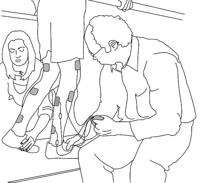
Sensory perception occurs when an organism is able to perform neurophysiological processing of stimuli in their environment and involves "the senses" such as touch , smell and sight. We are going to focus on touch and sight in this essay. The ability of a person to feel painless stimuli in the existence or absence of motion can be easily gauged with the use of electrical stimulation on the skin.(see figure 1.) The personal strength of the suprathreshold stimulation is not varied during motion. Inequity of slight changes in the strength of the suprathreshold stimulus is also not changed by motion while, in the same subjects, finding thresholds were raised when the stimulated arm was in motion. The findings indicate that the increase of detection thresholds while in motion can be justified by masking. Active motion and Passive motion of the limb being stimulated increase detection threshold .The active motion having a marginally larger and more constant effect that the passive motion. Therefore, equally central feedback and peripheral feedback factors can simulate in reducing one’s capability to recognise a gentle stimulation during motion (Chapman et al, 1987).
|
Effects of training on motor and sensory function after injury
There has been extensive research into plastic changes in the central nervous system of mammals prompted by life-changing situations amputation of a limb. Although there has been much research in this field it is still unknown about the degree to which the original pathway keeps continuing function after a limb amputation. Studies have shown that central nervous system pathways established with amputated peripheral nerves contain at minimum a few sensory or motor functions.
Studies are currently being carried out to find out if it were possible for these functional networks to be enhanced or stronger if the subject trained and worked on perfecting movement over a period of time. To conduct this research, electrodes are inserted into bundles of split nerves belonging to subjects that are long term amputees. Then, differences in electrically erased feelings and free motor neuron action linked with a subject trying to move a phantom limb. Stimulation of a nerve frequently occurred in discrete, exclusive, sorted feelings of pressure or touch and a sense of joint position. No compelling differences in the amount of stimulation parameters needed to cause the feelings were detected. Furthermore, the subject amputees had the ability to better their free power of motor neuron activity, however, the amount and consistency of difference was comparable to that observed with training in non amputee subjects on normal motor tasks. this research shows that the central plasticity that is observed post amputation is probably due to unmasking, and not restoration of current synaptic connections (Dhillon et al, 2005).
Peripheral nerve amputation has been shown to produce changes in cortical sensory and motor representation in various mammalian species, including humans. Contradictory to the general idea that there is a fixed wiring mechanism, there is substantial findings cortical representation of the limbs and body is frequently adjusted in reaction to skills, behavior or activity (Chen et al, 2002). Different findings show that is is feasible to bond microelectrodes at a sub fascicular level to little bundles of motor and sensory neutrons (Branner and Normann, 2000).
Phantom Pain
Treating phantom pain and providing sensory feedback to an amputee from a prosthetic limb
Phantom Pain is an indirect dysfunctional result that can affect a person after undergoing an amputation of a limb. They feel sensations of pain or discomfort arising from the original limb. The causes are not fully understood, however, several processes are responsible. The sensations are understood to emerge from the missing limb, although, sensory receptors from other parts of the body trigger these "feelings". These sensations are not always related to pain, however, when the pain is present it can be get so agonizing that it becomes insufferable or debilitating.
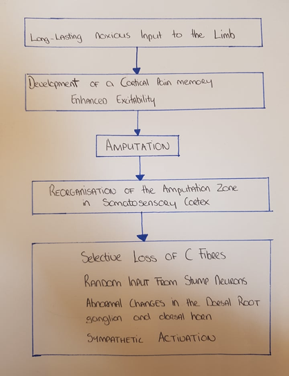
Amputation disrupts or eliminates the usual flow of sensory information originating from other processes of sensory receptors such as muscle receptors transported by large diameter, myelinated axons which usually transmit non-painful information of proprioceptive and cutaneous origin for example, pressure, muscle length, joint position or touch. (see figure 2.)
For transmission in the spinal cord and brain centers, the balance of activity in the large and small diameter sensory nerve fibers has become important. Fiber diameters of a severed nerve are reduced but the nerve cells continue to conduct electrical impulses and maintain their basic synaptic connectivity patterns (Hoffer et al, 1979). Sensory fibers degenerate more than motor fibers and the sensory fibers with small-diameters degenerate less than the large-diameter sensory fibers. "For hind limb nerves of cats that were cut and ligated over a period of 300 days, found that large sensory fibers had a 60% decrease in conduction velocity (CV); small sensory fibers had about a 45% decrease in CV; large motor fibers had a 40% decrease in CV: and small motor fibers had about a 20% decrease in CV. Thus, in amputated nerves, “large” and “small” nerve fibers will gradually become closer in their diameters and consequently closer in their thresholds for electrical stimulation." (Milner and Stein, 1981).
|
Their have been many propositions for the treatment of phantom limb pain, for example, antidepressant medication but they have severe side effects. Currently, there is no drug that can effectively treat these pains without side effects. The removal or blockade of sympathetic supply to the stump provides a reduction of phantom pain, however, it only has a temporary effect and it depends on how long after the amputation has been carried out (Livingston, 1945).
There is currently an invention that replaces lost sensory function and that eases phantom pain. Central perception of pain information transported by sensory fibers with a small diameter can be suppressed with the help of activity flowing centrally along fibers with a larger diameter. There is a greater likelihood of pain sensations reaching consciousness when there is a lack of activity that would usually happen in sensory fibers with larger diameters, for example, touch receptor afferentsthat detached from their peripheral sensory organ. When this condition occurs in amputees, selective electrical stimulation of the large diameter sensory fibers may reverse it by rectifying the excessive flow of pain information in central pathways by reinforcing a normal balance in small and large sensory fibers actions. The method involved implanting electrodes in or around the peripheral nerve stumps remaining within the proximal stump. The chosen electrical stimulation parameters provide sensory feedback about parameters of a prosthetic limb, for example touch and joint position and provides treatment of a phantom limb pain. The electrodes are incorporated within a tubular nerve cuff designed to be implanted in the limp stump so that it circumferentially surrounds a portion of the nerve (Hoffer, 1999).
Advances and improvements
Advances in medicine and intracortical micro stimulation
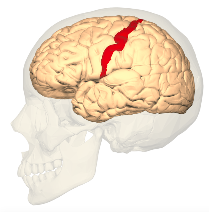
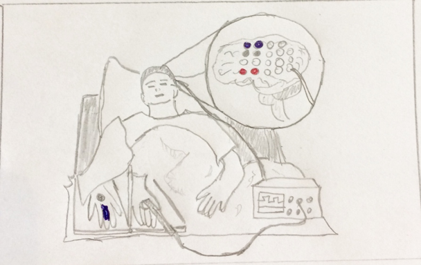
The Challenge is restoring the sense of touch in artificial limb prostheses. The difficulty is trying to regain the ownership of the limb and to be able to feel certain stimuli i.e., heat, pressure on the artificial limb. Previous research has been completed using a rubber hand illusion. The experiment involved the participants real hand (which was hidden) and also an artificial hand which was in the participant’s sight. During the experiment the fake hand was continuously touched with a probe (Botvinick and Cohen, 2008). This can activate ownership of the artificial hand in upper limb amputees. (Ehrsson et al, 2008) However, in patients with no peripheral stimulation due to injury of the spinal chord or nerves, it is difficult to prompt ownership when studies depend on touching stimulation and it is difficult to have the feeling as if the limb is their own as previous studies depend on touching of the real hand or on tactile stimulation of the stump (Ehrsson et al, 2008 and Schmalzl et al, 2011). It is possible to display sensory feedback from a robotic arm by administrating the feedback through electrodes implanted in the primary somatosensory cortex. (See figure 3.) (O’Doherty et al, 2011).This was done to try and bypass the Peripheral nervous system. The illusion of ownership of the rubber hand was induced using electrical brain stimulation of the hand primary somato sensory cortex. As a control, areas not around the hand where touched and this did not evoke the illusion. These findings show that the brain is able to integrate electrical cortical-somatosensory and natural visual signals to make coherent multisensory representations of one’s own limbs according to the same key rules of spatiotemporal congruence that carry out the normal perception of our body (Makin et al, 2008).
|
A digital probe was connected to a cortical stimulation device. The finger of the rubber hand was touched with the probe. This brought an electrical current across the 2 electrodes situated in the participants primary somato sensory cortex (blue electrodes in the figure). In the control the participant’s wrist was used. The current was brought to a pair of electrode’s partnered with somatosensation on the wrist (grey electrodes) The real hand of the participant was never touched. The red electrodes indicate a control stimulation area. (See figure 4.) Object manipulation depends on sensory signals. Somatosensory feedback is vital for the manipulation of objects in amputees. Advances have been made in trying to convey this sensory information needed for object manipulation. This has been done using intracortical microstimulation of somatosensory cortex. Experiments have been completed showing that we can evoke stimuli that are placed on localized patches of skin that track stimuli such as pressure and that signal the timing if contact events (Richer et al, 1993).
|
A major challenge in developing approaches to transmit sensory feedback using intracortical stimulation in animal models is to be able to analyse the evoked sensation (Schiller et al, 2011).Animals can be thought to discriminate sensory stimuli along a dimension of interest and then see if the animal can perform the task when physical stimuli are replaced with intracortical microstimulation (Romo et al, 1998 and Romo et al, 2000). Intracortical microstimulation systems are made to mimic the patterns of neuronal activation that encode the sensory dimension. contact location, pressure, and timing are three of the simplest cutaneous signals that bring about object grasping and manipulation (Johansson and Flanagan, 2009). The somatotopic arrangement is very important as the neural coding of the stimulus area depends on this.
The regulation of where on the body the sensation is felt is dependent on the activated neurons within the body representations in the primary somatosensory cortex. (Rasmussen and Penfield, 1947). Information can be conveyed about location by targeting intracortical microstimulation on populations of neurons with specific receptive field (RF) locations. There are mechanisms such as 1. when the pressure on the skin increases, the neuronal population with Rfs under stimulus is more active and 2. neurons with RFs next to them will be activated so the activated neuronal population will increase (Simons et al, 2005). information can be displayed about pressure by increasing the amplitude of intracortical microstimulation so both the strength of activation of neurons near the electrodes and the size of the activated population can be increased (Tehovnik et al, 2006). The neural coding of contact timing—which signals when contact with an object is initiated and terminated—is thought to depend on the on and off responses made in primary somatosensory cortex neurons at the onset and offset of contact (Pei et al, 2009). These temporally precise responses are relatively insensitive to object properties (Bensmaia et al, 2008) and critical in guiding the manipulation of objects (Rasmussen and Penfield, 1947). information may be shown about contact timing by delivering phasic Intracortical microstimulation at the onset and offset of object contact. An experimental approach consists in mimicking natural patterns in the brain and monitoring whether the animal spontaneously interprets these evoked patterns in the right way.
Conclusion
In conclusion, it is important to block phantom pain because after long term inability to physically touch or treat the sensation it would have severe psychological consequences however the advances in manipulating artificial sensoring means that amputees will have a varied sense of feeling. In the future we would like to see more improvements made in attempting to feel varied textures such as rough vs smooth, dull vs sharp or wet vs dry and stimuli like heat, pressure and temperature.
Bibliography
References
- Bensmaia, SJ.; Denchev,PV.; Dammann, JF.; 3rd, Craig, JC.; Hsiao, SS. (2008): The representation of stimulus orientation in the early stages of somatosensory processing.J Neurosci 28(3):776–786.
- Botvinick, M.; Cohen, J. (2008): Rubber hands ‘feel’ touch that eyes see. Nature. 1998;391(6669):756.
- Branner, A.; Normann, RA. (2000): A multielectrode array for intrafascicular recording and stimulation in sciatic nerve of cats. Brain Res Bull 51: (4) 293 - 306.
- Chapman, CE.; Bushnell, MC.; Miron, D.; Duncan, GH.; Lund, P. (1987): Sensory Perception During Movement in Man. Experimental Brain Research 68: (3) 516-524.
- Chen, R.; Cohen, LG.; Hallett, M. (2002): Nervous system Reorganisation Following Injury. Neuroscience 111: (4) 761 - 73.
- Dhillon, GS.; Kruger, TB.; Sandhu, JS.; Horch, KW. (2005): Effects of Short-Term Training on Sensory and Motor Function in Severed Nerves of Long-Term Human Amputees. Journal of Neurophysiology 93: (5) 2625-33.
- Ehrsson, HH.; Rosén, B.; Stockselius, A.; Ragnö, C.; Köhler, P.; Lundborg,G . (2008): Upper limb amputees can be induced to experience a rubber hand as their own. Brain. ;131(Pt 12):3443–3452
- Hoffer, JA.; Stein, RB. and Gordon, T. (1979): Differential Atrophy of Sensory and Motor Fibers Following Section of Cat Peripheral Nerves. Brain Res. 178:347-361.
- Johansson, RS.; Flanagan, JR. (2009): Coding and use of tactile signals from the fingertips in object manipulation tasks. Nat Rev Neurosci 10(5):345–359.
- Livingston, KE. (1945): Phantom Limb Syndrome. A Discussion of the Role of Major Peripheral Nerve Neuromas. J. Neurosurgery 2:251-5.
- Makin, TR.; Holmes, NP.; Ehrsson, HH. (2008): On the other hand: Dummy hands and peripersonal space. Behav Brain Res.;191(1):1–10.
- Milner, TE.; Stein, RB. (1981): The Effects of Axotomy on the Condition of Action Potentials in Peripheral Sensory and Motor Nerve Fibres. J Neurol Neurosurg Psychiatry 44(6):485-96.
- O’Doherty, JE.; Lebedev, MA.; Lfft, PJ.; Zhuang, KZ.; Shokur, S.; Bleuler, H.; Nicolelis, MA. (2011): Active tactile exploration using a brain-machine-brain interface. Nature. ;479(7372):228–231.
- Pei, YC.; Denchev, PV.; Hsiao, SS.; Craig, JC.; Bensmaia, SJ. (2009): Convergence of submodality-specific input onto neurons in primary somatosensory cortex. J Neurophysiol 102(3): 1843-1853.
- Rasmussen, T.; Penfield, W. (1947): The human sensorimotor cortex as studied by electrical stimulation. Fed Proc6(1 Pt 2): 184.
- Richer, F.; Martinez,M.; Robert, M.; Bouvier, G.; Saint-Hilaire,JM. (1993): Stimulation of human somatosensory cortex: Tactile and body displacement perceptions in medial regions. Exp Brain Res 93(1):173-176.
- Romo, R.; Hernández, A.; Zainos, A.; Brody, CD.; Lemus, L. (2000) : Sensing without touching: Psychophysical performance based on cortical microstimulation. Neuron 26(1):273-278.
- Romo, R.;Hernández, A.; Zainos, A.; Salinas, E. (1998): Somatosensory discrimination based on cortical microstimulation. Nature 392(6674):387-390.
- Schiller, PH.; slocum,WM.; Kwak, Mc.; Kendall,GL.; Tehovnik,EJ. (2011): New methods devised specify the size and color of the spots monkeys see when striate cortex (area V1) is electrically stimulated. Proc Natl Acad Sci USA 108(43):17809-17814.
- Schmalzl, L.; Thomke, E.; Ragnö, C.; Nilseryd, M.; Stockelius, A.; Ehrsson, HH. (2011): "Pulling telescoped phantoms out of the stump": Manipulating the percieved position of phantom limbs using a full-body illusion. Front Hum Neurosci.; 5:121.
- Simons, SB.; Tannan, V.; Chiu, J.; Favorov, OV.; Whitsel, BL.; Tommerdahl, M. (2005): Amplitude-dependency of response of SI cortex to flutter stimulation. BMC Neurosci6:43.
- Tehovnik, EJ.; Tolias, AS.; Sultan, F.; Slocum, WM.; Logothetis, NK. (2006): Direct and indirect activation of cortical neurons by electrical microstimulation. J Neurophysiol 96(2):512-521.
Other:
- Hoffer, JA. (1999): Electrical stimulation system and methods for treating phantom limb pain and for providing sensory feedback to an amputee from a prosthetic limb. Neurostream Tech Inc.
Figure references
Figure 1. "electrical stimulation on skin" https://upload.wikimedia.org/wikipedia/commons/5/55/Functional_Electrical_Stimulation_Therapy_for_improving_walking_in_incomplete_spinal_cord_injured_individuals_-30%2C31-.jpg
- Figure 2. "The main factors thought to be relevant to phantom limb pain" - by Hannah Kelly
Figure 3. "The primary somatosensory cortex of the human brain" - Body Parts 3D/Anatomography - https://upload.wikimedia.org/wikipedia/commons/e/e6/BA312_-_Primary_Somatosensory_Cortex_-_lateral_view.png
- Figure 4. "Experimental set up of cortical stimulation device" - by Emma Cummins


